Download
1 / 9
100 likes | 235 Views
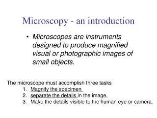
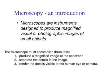
Microscopy - an introduction. Microscopes are instruments designed to produce magnified visual or photographic images of small objects. The microscope must accomplish three tasks 1. produce a magnified image of the specimen separate the details in the image,

E N D
Microscopy - an introduction • Microscopes are instruments designed to produce magnified visual or photographic images of small objects. • The microscope must accomplish three tasks • 1. produce a magnified image of the specimen • separate the details in the image, • render the details visible to the human eye or camera.
Electron Microscopes • The use of high energy electrons to examine the fine details of objects.
Major Types of electron microscopes. • Scanning electron microscope (SEM). • Transmission electron microscope (TEM)
Scanning electron microscopy (SEM). • Types of specimens: • -Whole organisms • -Natural tissue surfaces • -Exposed tissue structure • -Corrosion casts
Transmission electron microscopy (TEM). • Allows the observation of molecules within cells • Allows the magnification of objects in the order of 100, 000’s.
Representative EM images of Pst DC3000 avrPto (pAVRPTO), immunogold-labeled with the AvrPto antibody. In situ immunogold labeling was done after bacteria were grown in hrp-inducing medium for 4 hours, supplemented with (A) no SA, (B) SA for 4 hours, or (C and D) SA for 1 hour. Dark dots are 15-nm gold particles in (A) and (C) and 10-nm gold particles in (B) and (D). Arrows indicate Hrp pili attached to rod-shaped bacteria (only a portion of the bacterium is shown, surrounded by dark stain). Scale bars, 100 nm.