Download

1 / 85
890 likes | 1.23k Views
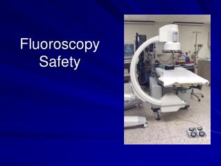
The Safe Use of Fluoroscopy. A required inservice for people who work with or near fluoroscopy. The FDA says . . . .

E N D
The Safe Use of Fluoroscopy A required inservice for people who work with or near fluoroscopy
The FDA says . . . “The Food and Drug Administration Center for Devices and Radiological health has received reports of occasional, but at times severe, radiation-induced skin injuries to patients resulting from prolonged, fluoroscopically-guided, invasive procedures. Physicians performing these procedures should be aware of the potential for serious, radiation-induced skin injury caused by long periods of fluoroscopy during these procedures.” from FDA Public Health Advisory, Sept. 30, 1994
The State of Ohio says . . . “All individuals operating fluoroscopic equipment, and individuals likely to receive an annual effective dose equivalent in excess of 1 mSv from participating in fluoroscopic procedures, shall receive at least two hours of radiation protection training specific to fluoroscopy in addition to [other required training]. . . prior to performing or participating in fluoroscopic procedures.” Ohio Administrative Code 3701:1-66-07 (G)
The Kettering Health Network says. . . All persons operating or working directly with fluoroscopy equipment at any KHN facility must have proof of taking this inservice or an equivalent one at any other facility within the State of Ohio.
The Good News • This inservice is only required once before working with fluoroscopy. • (A high badge reading may require that you take a one-hour refresher course.) • This inservice is good anywhere in the State of Ohio. • If you have done a similar inservice at another facility within Ohio, provide the Radiation Safety Office with documentation, and you are excused from this one.
One Little Detail • While this training is good for each hospital where you work, it must be approved by the Certified Radiation Expert (CRE) for that hospital. • You may need to provide a copy of this presentation to the CRE.
Who’s Who • The Ohio Department of Health, Bureau of Radiation Protection is responsible for enforcing rules concerning the safe use of radiation, including fluoroscopy. • Information on how to contact them is posted in every radiation use area throughout the network. • They are also on-line at www.odh.ohio.gov.
Who’s Who • The Kettering Health Network Radiation Safety Office is responsible for overseeing radiation safety throughout the entire Network. • They are located on the ground floor, NW wing of Kettering Hospital. • They have a site on the KHN intranet (which may be where you are viewing this presentation.)
The Radiation Safety Office • Radiation Safety Officer Steven Cartwright, PhD, DABR, DABMP 395-8818, pager 370-0006 • Radiation Safety Specialist Mark Berner, MBA 298-3399, X57704, pager 370-0005
Who else is Who • The Food and Drug Administration (FDA) is responsible for the safe design of X-ray units, including fluoroscopes. • They do not govern the use of fluoroscopes, but in 1994 they alerted the medical community to the possibility that fluoroscopy can be misused and cause really ugly skin burns to patients. • Some of those pictures will show up later in this presentation.
Some Ohio basics • In the State of Ohio only licensed operators can use a fluoroscope on humans. • Physicians are specifically exempted from needing a license. They can use a fluoroscope on anybody as long as it’s within the scope of their practice. • NO NURSES! (Unless they hold a radiographer’s license, in which case they are RTs as well.) • THE PHYSICIAN IS ALWAYS RESPONSIBLE FOR APPLYING RADIATION TO A PATIENT, NO MATTER WHOSE FOOT IS ON THE PEDAL.
What Are X-rays Anyways? • X-rays are high-energy electro-magnetic waves generated by machines. • X-rays are exactly the same as visible light, only with 30,000 times more energy. • Because of their high energy X-rays can penetrate tissue, but they aren’t so good with bone. • The difference in penetration makes up the classic X-ray image.
The Classic X-ray Image • The first X-ray image taken by Wilhelm Roentgen. • Incidently, that’s Mrs. Roentgen’s hand. You didn’t think Wilhelm would expose himself, did you?
An X-ray Myth • X-rays do not stay in the body. • They don’t make you radioactive. They don’t make you glow. • When you turn off the X-ray machine they’re gone, just like the light from a light bulb. • People who have had an X-ray procedure don’t pose any special threat to the rest of the world. • Unless they text while driving.
Fluoroscopy • Fluoroscopy is real-time X-ray imaging. • Instead of a piece of film, the fluoroscope image is formed on an image intensifier (the “II”). It is displayed on a TV monitor.
Types of Fluoroscopes • General purpose fluoroscopes may have the X-ray head under or over the table. • Over-table units can be used for general radiology as well. • The II on an under-table unit is called the tower for an obvious reason.
Stationary Fluoroscopes • Undertable • Over table X-ray head Image intensifier
Types of Fluoroscopes • If the X-ray head and II are linked together by a large C-shaped arm the fluoroscope is a called a C-arm. • Mobile C-arms can roll around. Mini C-arms are tiny fluoroscopes used only for extremities.
C-arms • Mobile C-arm • Mini C-arm
Proper Positioning • Whenever possible, position the X-ray head under the patient. (This isn’t possible on an over-table unit.) • The X-ray source (inside the head) must be at least 12” away from the patient’s skin. This is built in to most fixed units. C-arms have spacer cones to keep the right distance.
Foot Pedals • All fluoroscopes of any type are operated by a foot pedal. • The pedal is a “dead man” switch. The machine will only operate when there is pressure on the switch. • Presumably if you die while operating a fluoroscope you will release the foot pressure.
Image Intensifiers • The Image Intensifier (II) converts the radiographic image into a TV image and makes it considerably brighter. • The II may be a large vacuum device or a flat panel. • II’s can magnify the image.
Image Intensifiers Flat Panel Tube
The Fluoroscopic Image • Compared to conventional X-ray images, fluoroscopic images are grainy. • This is because fluoro images use as little radiation as possible. • Less radiation = more noise (image grain) • More noise = harder to see fine detail
Image Noise • The grainy appearance of an image is called noise. • It’s just like static on the radio. • The main cause of noise is too little radiation. • Too much radiation makes a great image but overdoses the patient—and you.
The Effect of Noise • The image is the same each time, but the noise increases. • In the noisiest image (right) the dim circles are still there, but you can’t see them. • This is like fluoroscopy.
Why this is OK • Fine image resolution is needed to diagnose unknown conditions. You need to see everything. • Fluoroscopy is rarely used for diagnosis. • Under fluoro you’re usually looking for something large or filled with contrast, and sometimes both.
Physics Alert! Formal discussion of X-ray imaging to follow
Generating X-rays • X-rays are generated in a vacuum tube. • Electrons from a cathode accelerate under high voltage towards an anode. • When the electrons hit the anode they slow down abruptly and generate X-rays in the process. • The X-rays escape out a window in the tube and become useful.
X-ray Technique • The energy of the X-rays is determined by the voltage applied to the anode (commonly called the kilovoltage or kV). • The intensity of the X-rays is determined by the flow of electrons from the cathode to the anode. • The electron flow is often called the mAs (for milliamp-second). • Together the kV and mAs make up the X-ray technique.
X-ray Technique—kV • The kV determines how much energy gets through the subject. • Use high kV to penetrate bone, large people, or large boney people. • Use low kV to show soft tissues.
X-ray Technique—kV • kVs range from about 25 kV for mammography to 150 kV for chest X-rays. • Typical fluoroscope kVs are in the 50 kV to 80 kV range.
X-ray Technique—mAs • The mAs determines the total amount of radiation used to make an image. • In a conventional X-ray the time (s) is very short and the tube current (mA) is fairly large. • Fluoroscopy uses a small mA, but exposures may run for several seconds. • The result is a moving, but grainy image.
X-ray Technique—kV and mA • Typically, the kV is set to penetrate the chosen body part, and the mA is adjusted to give a suitable image. • In fluoroscopy this procedure is usually done automatically in a process called auto-fluoro.
Auto-fluoro • Step on the pedal and the machine automatically adjusts the kV and mA to give a good image. • As you move the fluoroscope around the kV and mA automatically adjust. • You may not know at any moment just what the kV and mA are.
Pulsed Mode • Fluoroscopy often uses pulsed X-rays. • The radiation is emitted in small bursts from 2 to 30 times per second. • Anything higher than 30 times per second looks like a continuously moving image to the human eye. • At slower rates the image may look jerky, but it is still useful for most tasks.
Patient Dose • Only a small portion of the radiation from a fluoroscope makes it to the II. • The rest stays in the patient. This energy is the patient dose. • The patient dose is responsible for damage to a patient’s skin or increase in cancer risk. • More about this later. Just keep reading.
The Terms We Use • Three different terms are used to describe the interaction of radiation with the human body (well, anything, actually). • Exposure—The “amount” of ionizing radiation present at a point in space • Dose—The amount of energy absorbed in a mass of tissue • Effective Dose—A derived quantity that describes the biological effect of absorbed radiation • These three concepts are closely related, but measured by different units with slightly different meanings.
The Units We Use • There are two sets of units used to describe radiation—older ones, often found in textbooks, and newer ones that conform to the conventions of the System Internationale. • The relation between them is sort of like the relation between currencies. Two kinds of currency both describe the value of something, but they use different numbers and units. (Anybody who ever had to work with the old British system of pounds, shillings, and pence can appreciate this.)
The Units We Use(the m before a unit stands for milli-, or 1/1000th) Old Units • Exposure—Roentgen (R, mR) • Dose—rad (r, mrad) • Effective dose—rem (rem, mrem) New Units • Exposure—Air Kerma (no accepted abbreviation yet) • Dose—Gray (Gy, mGy) • Effective dose—Sievert (Sv, mSv)
Where You Find These Terms • The radiation output of an X-ray machine is usually described by the exposure rate, R/min, measured at a fixed point in front of the X-ray head (usually 1 meter or 40 inches). • The dose (sometimes called organ dose) is calculated to determine the effect of radiation on a particular organ. This is used a lot in radiation therapy treatment planning. • The effective dose is calculated from the exposure of a film badge and reported as part of your monthly badge reading.
Biological Effect of Radiation • Radiation ionizes (knocks off electrons from) atoms. • In the human body, mostly it ionizes water. • The ionized water breaks up into radicals that break DNA chains.
The Mysteries of DNA • If left alone long enough, a broken DNA chain can repair itself. No harm done. • If the cell divides before the chain repairs itself, the cell dies. • Enough cells die and bad things happen. • Cells die when they divide too fast to repair themselves or when the radiation is coming in too fast to give them a chance.
Rapidly dividing cells (most sensitive) sperm lymphocytes small intestine stomach Slowly dividing cells (least sensitive) bone skin nerves colon Radiation Sensitivity
The Nature of Radiation Damage • Radiation damage seldom appears at the time of irradiation. • Visible effects occur later, from several hours to nearly a year. • The first (almost) visible effect is a drop in the white blood count. • The first external visible effect is a skin burn.
A Note About Radiation Therapy • Cancer cells divide much more than normal cells. • Radiation kills both normal and cancer cells, but the cancer cells have less opportunity to repair themselves. • Repeated applications of radiation eventually kill all of the cancer cells while the normal cells recover.
Why This is Important • Diagnostic X-ray procedures do not usually use enough radiation to cause damage. • Fluoroscopy is the one modality capable of delivering sufficient energy to one spot so as to cause damage.* • Educating the operators can prevent damage from occurring. • *Recently a CT was used to burn people. Who knew it was possible?