Download
1 / 87
920 likes | 2.05k Views
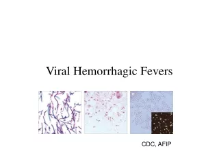
Viral Hemorrhagic Fevers. The Filoviruses Steve Vivian Jenn. Live Footage. Ebola Virus Outbreak. Viral Hemorrhagic Fevers. Severe multi-system syndrome (multiple organs affected). Vascular system is damaged and body loses the ability to regulate itself. Accompanied by hemorrhaging.

E N D
Viral Hemorrhagic Fevers The Filoviruses Steve Vivian Jenn
Live Footage Ebola Virus Outbreak
Viral Hemorrhagic Fevers • Severe multi-system syndrome (multiple organs affected). • Vascular system is damaged and body loses the ability to regulate itself. • Accompanied by hemorrhaging. • Many VHF viruses cause life threatening diseases • Most have no established treatment or cure.
VHF Caused by Viruses of 4 Families • Arenaviruses • Filoviruses (Ebola and Marburg) • Work with filovirus species requires Biosafety Level 4 (BSL-4) “space suit” containment (HIV requires BSL-2 “space suit” containment). • Bunyaviruses • Flaviviruses
Features of these Viruses • RNA Viruses, covered in lipid coating • Humans are not natural reservoir, but can transmit virus • Viruses are restricted to areas of their host species
Marburg • Caused by a zoonotic virus of the filovirus family. • Mortality Rate 23-25%
First recognized in 1967 when simultaneous outbreaks occurred in Marburg and Frankfurt, Germany, and in Belgrade, Yugoslavia. • 37 people became ill. The ill were mostly laboratory workers and medical personnel. • Original people who became ill had been exposed to the tissues of African green monkeys, which were imported from Uganda for research.
Location: • The area to which the virus is native is unknown, but it is believed to include parts of Uganda, Western Kenya, and Zimbabwe. • Transmission • The animal host to the Marburg Virus is unknown, and so is the way that the animal transmits the disease to humans. • People who have been exposed to infected monkeys or their body fluids have become infected in the past.
Transmission (continued) • The disease is easily transmitted between humans. Direct contact with an infected person, or exposure to their body fluids, are both ways by which the disease is thought to be transmitted.
Ebola • Believed to be caused by a zoonotic member of the filovirus family. • Believed to be carried by an animal host that is native to Africa. • Named after a river in the Democratic Republic of Congo in Africa (formerly Zaire) where first outbreak occurred in 1976. • First Ebola outbreak in Zaire (1976)- 318 Human cases, 88% of those people died of the disease.
Great Plague of Athens529 B.C. • Thucydides wrote about a disease with symptoms that are very similar to Ebola, including: • High Fever, Vomiting, Chest Pain, Diarrhea, fetid breath and a bumpy red rash.
4 Types of Ebola Virus • Ebola-Zaire • Ebola-Sudan • Ebola-Ivory Coast • Ebola Reston All of these strains have been known to cause disease in humans, except for Ebola-Reston, which has only caused disease in Non-Human Primates. Electron Micrograph of Ebola Virus
Location: • The Ebola Virus has been reported in the Democratic Republic of Congo, Gabon, Sudan, the Ivory Coast, and Uganda. Map of Ebola Outbreaks in Africa
Ebola-Reston outbreak at primate research facility in Virginia. Virus was carried by monkeys that had been imported from the Phillipines. • 4 humans were infected and developed antibodies, but none of them became sick Map of Ebola Reston Infected Monkeys in Philippines
Transmission • It is believed that the first human becomes infected with Ebola through contact with an infected animal. • An outbreak of Ebola-Zaire occurred in Gabon in 1996 after people had eaten an infected monkey. • One Scientist who had conducted an autopsy on a wild chimpanzee in the Tai Forest in Ivory Coast was diagnosed with Ebola-Ivory Coast in 1994.
People can become infected with Ebola if they come into contact with the blood or secretions of an infected person. • People can also become infected through contact with objects, such as needles, that are contaminated with secretions or blood. • In 1976, in England, a person became infected with Ebola-Sudan after being accidentally pricked with a contaminated needle.
Because the disease is easily transmitted, Nurses must wear protective clothing when interacting with a patient and must dispose of all needles and other medical supplies properly. Nurses in full protective gear in Gabon, 2002
Patients who have died of the disease must be buried properly. Care must be taken that there is no contact with the deceased patient. Burying Ebola Victim in Gabon, 2002
The Ebola Virus takes a psychological toll as well, causing devastated villagers to search for scapegoats. Posted, February 21, 2003 8:37 AM EST Brazzaville, Congo- Congolese villagers have stoned and beaten to death four teachers accused of casting an evil spell to cause an outbreak of the deadly Ebola disease that killed nearly 70 people, a local official said Friday. (Reuters)
Relief , joy and celebration after the Gabonese Health Ministry declares that the Ebola outbreak has ended. (May 6, 2002)
Pleomorphic • Definition: having the ability to assume different forms or shapes • Four main shapes: long filamentous, U shaped, 6 shaped, circular
Structure • Filoviruses are non-segmented negative strand • viruses • Composed of a helical ribonucleoprotein complex • (nucleocapsid) • Covered with a host-cell derived membrane envelope • Matrix between the envelope and nucleocapsid is • filled with a protein lattice
Viral Genome • Consists of seven genes • These genes encode seven proteins in the Marburg virus and eight in the Ebola virus • Both viruses have a 3’ leader and 5’ trailer sequence that is highly conserved and has a high degree of complementarity • These extragenic seguences are important in the initiation of transcription and replication
Viral Proteins * Ebola virus only • NP: Nucleoprotein – the primary structural protein associated with the filovirus nucleocapsid
Viral Proteins * Ebola virus only • VP35: acts as a cofactor in transcription and replication of the viral proteins
Viral Proteins * Ebola virus only • VP40: a matrix protein, also the most abundant viral protein; may facilitate in the budding process
Viral Proteins * Ebola virus only • GP: Glycoprotein – makes up the virion spikes or peplomers and mediates entry into host cells through receptor binding
Viral Proteins * Ebola virus only • sGP: secreted from the cell, present in large amounts in the blood of Ebola victims; may help to inhibit the immune response
Viral Proteins * Ebola virus only • VP30 – a minor nucleoprotein that may be involved in securing the RNA to the C terminus of NP
Viral Proteins * Ebola virus only • VP24: unknown function, possibly a matrix protein
Viral Proteins * Ebola virus only • L: Polymerase L – acts as the polymerase and is the largest and least abundant viral protein
Entrance into Cells • Mediated by the Glycoprotein on the envelope surface
Structure of GP • Precursor form GP0 is cleaved yeilding GP1 and GP2, they are connected via one disulfide bond • Form a heterodimer • Peplomers are then formed by the association of three of these heterodimers to form a homotrimer • GP2 is responsible for trimerization, fusion of the envelope and cell, and has an immunosuppressive motif • GP1is the receptor binding domain
Receptor - Lipid Rafts • Receptor, while for most cells unknown, appears to be located in LIPID RAFTS : less fluid areas of the membrane high in cholesterol and glycosphingolipid; the cholesterol-binding caveolin is also closely associated with the rafts
Endocytosis • Caveolae are present in the plasma membranes of most cell types and are thought to form from lipid rafts • The major structural protein is caveolin, a multipass protein • Caveolae invaginate and collect their cargo proteins, but do not require the assembly of a cytosolic protein coat
Transcription • Takes place in cytoplasm of cell • The seven genes are transcribed to produce seven monocistronic polyadenylated mRNA’s • Each gene has a conserved transcriptional stop and start sequences • Transcripts are thought to have 5’ caps • Gene overlaps - function unknown
Unusual GP Transcription • In the Ebola Virus the GP gene codes for GP and sGP • sGP is the primary gene product • Full length GP is expressed by transcriptional editing of a single adenosine at a run of seven uridine residues in the genomic RNA
Translation • sGP and GP are translated by membrane bound ribosomes and enter the ER and follow the exocytotic transport route to the cell surface • All other viral proteins are translated by free ribosomes in the cytosol
Replication • Build up of proteins signals switch to replication • Switch results in synthesis of + sense RNA as a template • Depletion of proteins causes switch back to transcription, eventually equilibrium
Immune System Evasion • Host dies with little evidence of an immune response • Mitosis of lymphoid cells seen to decrease 2-3 days postinfection with eventual apoptosis • Swelling of MPS and stromal cells and eventual lysis • Dendritic (antigen-presenting) cells also undergo apoptosis
Immune System Evasion • sGP may bind to neutrophils and inhibit their activation • sGP as a decoy? – absorb neutralizing antibodies • VP35 may act as an IFN antagonist • GP2 may have an immunosuppressive role • Ebola shown to inhibit induction of immunomodulatory and antiviral genes in endothelial cells (MHC I, IFN, etc…)
Cytotoxicity,Hemorrhage, and Shock • GP expression causes downregulation of the cell-surface expression of integrins – cell rounding and detachment seen • Disruption of cell functions in general • Release of vasoactive agents (cytokines, chemokines, histamines and peroxidases) from infected monocytes and endothelial cells could increase hemorrhaging and induce shock
Risk Factors • Travel to Asia or Africa • Handling of animal carcasses • Contact with sick animals or people • Arthropod bites within 21 days of onset of symptoms • Transmission is highest during latter stages of illness.