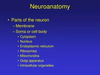
Functional Neuroanatomy
Functional Neuroanatomy. Next week’s reading…. STRUCTURE AND FUNCTIONS OF CELLS OF THE NERVOUS SYSTEM *CARLSON, N. R. XRX.57772 SCOTT SCOTT-RESV (Available: 2HOUR) . Overview. A word about the funny names, and a few definitions Anatomy versus Function: The Triune Brain Hypothesis


Functional Neuroanatomy
E N D
Presentation Transcript
Functional Neuroanatomy Functional Neuroanatomy
Next week’s reading… STRUCTURE AND FUNCTIONS OF CELLS OF THE NERVOUS SYSTEM *CARLSON, N. R. XRX.57772 SCOTT SCOTT-RESV (Available: 2HOUR) Functional Neuroanatomy
Overview • A word about the funny names, and a few definitions • Anatomy versus Function: The Triune Brain Hypothesis • The Brainstem • The Limbic System • Cerebral Cortex Functional Neuroanatomy
A word about the funny names… • Early anatomists named most brain structures (in Latin, for the most part) according to their similarity to commonplace objects: • amygdala = almond, • hippocampus = sea horse, • genu = knee, • cortex = bark • pons = bridge Functional Neuroanatomy
A few definitions Locations in the Brain • Described relative to neuraxis - an imaginary line drawn through the spinal cord up to the front of the brain • The front end is anterior • The back end is posterior • The terms rostral (toward the head) and caudal (toward the tail) are also used when referring to parts of the brain Functional Neuroanatomy
A few definitions • Dorsal (back) refers to the top of the head and the back • The ventral (front) surface faces the ground. • These directions are somewhat more complicated in humans because our neuraxis bends, so that the top of the head is now perpendicular to the back. Functional Neuroanatomy
The Neuraxis Dorsal Anterior Posterior Ventral Functional Neuroanatomy
Dorsal Anterior Dorsal Ventral Posterior The Neuraxis Functional Neuroanatomy
A few definitions • Lateral means to the side (away from the neuraxis) • Medial (or mesial) means toward the middle (towards the neuraxis) • Proximal: Areas of the brain that are near to one another • Distal: Areas that are far from one another Functional Neuroanatomy
A few definitions • Ipsilateral refers to structures on the same side of the body • E.g. the olfactory bulbs send ipsilateral connections to the brain - the right bulb connects to the right hemisphere, and the left bulb connects to the left hemisphere • Contralateral refers to structures on opposite sides of the body • E.g. the left motor strip connects to the right side of the body Functional Neuroanatomy
A few definitions Slices of the brain • Transversely, like a loaf of bread - also called frontal or cross sections • Parallel to the ground, giving us horizontal sections • Perpendicular to the ground and parallel to the neuraxis - sagittal section - midsagittal plane divides the brain in half along the longitudinal fissure • Because of our upright posture, cross sections of our spinal cord are actually parallel to the ground Functional Neuroanatomy
Transverse/ Cross section Horizontal section Sagittal section Functional Neuroanatomy
Gross Anatomy of the Brain Functional Neuroanatomy
Gross Anatomy of the Brain • Functional divisions based on evolutionary criteria Triune Brain Hypothesis (MacLean) “In its evolution, the forebrain of advanced mammals has expanded as a triune structure that anatomically and chemically reflects ancestral commonalities with reptiles, early mammals, and late mammals.” (1985) • Mammalian brain structure reflects its phylogeny Functional Neuroanatomy
Gross Anatomy of the Brain The three brains… 1. Reptiles (R-complex) = Brainstem • Basic regulatory and vegetative functions, “instincts” 2. Early Mammals = Limbic Cortex • Limbus = border or margin (Latin) • Explicit memories, emotion • Care and protection of offspring • Establishing territory 3. Late Mammals = Neocortex • Abstract reasoning, long-term planning and behaviour, higher sensory functioning, etc. Functional Neuroanatomy
Is the Triune Brain Hypothesis Useful? • Intuitive, “textbook” way of dividing the structure of the brain based on its evolutionary usefulness and function • Most textbooks dealing with brain function use the triune division • Accounts how the mammalian brain evolved and became specialized, beyond basic survival functions common to all animals • Accounts for disproportionately large telencephalon Functional Neuroanatomy
Is the Triune Brain Hypothesis Useful? 2. Emphasizes a distributed “systems” approach • As opposed to a modular approach • Processing of information proceeds through increasing levels of complexity • Multiple brain areas accomplish complex tasks • Diversity/redundancy of systems (I.e. memory) • Explains why partial functioning or recovery is possible Functional Neuroanatomy
Is the Triune Brain Hypothesis Useful? 3. Competition between different brain areas • Conflict between different phylogenic areas of the brain--situations where different functions come into conflict. (I.e. smoking) • Antagonistic or inhibitory areas exist in the brain. • Mental disease may reflect an imbalance: over- or underactivity of a brain area • Role of drug or treatment is to restore balance Functional Neuroanatomy
The Brainstem • Consists of all structures from the thalamus to the spinal cord • Regulatory functions: Eating, drinking, body temperature, sleep and waking, basic movement and learning • Generally speaking, these structures rule functions that are hard-wired, automatic, and not very plastic Functional Neuroanatomy
The Brainstem Functional Neuroanatomy
The Brainstem • Thalamus: A relay centre for sensory information (touch, vision, hearing); located near the middle of the cerebral hemispheres. • Fibres project to primary sensory areas in neocortex • There are separate nuclei for vision, touch, hearing • Not a “passive” structure because the majority (80%) of its connections are not from sensory neurons, but from the neocortex (including motor areas). Functional Neuroanatomy
The Brainstem • Hypothalamus: Controls all aspects of motivated (pleasure and pain) and regulatory behaviour • Autonomic (vegetative) system • Superior to the pituitary gland, reciprocal connections with it • “Master gland”: Closely involved in the regulation and secretion of hormones • 0.3% of the brain’s weight Functional Neuroanatomy
The Brainstem • Reticular Formation: Constellation of 90+ nuclei at the base of the brainstem • Bundles of fibres as well as projections that pass through to the forebrain from the spinal cord • A host of regulatory vegetative functions • connections with cerebral cortex and thalamus Functional Neuroanatomy
The Brainstem • Cerebellum: conspicuous bulbous structure protruding from the posterior brain; “little brain” • Distinctive narrow folds (folia), similar to sulci in neocortex • Involved in aspects of learning and coordination of skilled or smooth movement • Posture, walking, equilibrium Functional Neuroanatomy
The Brainstem • By and large, there is little that distinguishes mammals from reptiles in terms of brainstem structure and function • An evolutionary turning point occurred with the specialization of limbic cortex in mammals • Functions of brainstem aren’t replaced; rather, they are modified or enhanced by interacting with newer phylogenic structures. Functional Neuroanatomy
The Limbic Cortex • Three-layer cortical structure covering the periphery of the brainstem, on the ventral surface of the lateral ventricles • Primarily known for its role in emotion, (emotional) learning and memory • Also plays a role in spatial learning and olfaction (memories of odour) Functional Neuroanatomy
The Limbic Cortex Functional Neuroanatomy
The Limbic Cortex • Hippocampus: Located next to the lateral ventricle in the temporal lobe. • Along with the fornix, mammillary bodies, and cingulate gyrus is involved in learning and memory. • Amygdala: located anterior to the hippocampus • only part of the limbic system responsible for emotional responses • Other areas involved with learning and memory of emotions (recognition of emotional events) Functional Neuroanatomy
The Limbic Cortex Fornix: a bundle of axons that connects hippocampus with other regions of the brain, including the mammillary bodies (containing some of the hypothalamic nuclei) Functional Neuroanatomy
Cerebral Cortex • Six-layered structure • Includes most of the two symmetrical cerebral hemispheres. • The cerebral hemispheres contain the limbic cortex • The majority of the surface of the cerebral hemispheres is called the neocortex • Part of the cerebral cortex is buried in the frontal lobes (I.e. insula – taste, sensation, and memory) Functional Neuroanatomy
Cerebral Cortex • In humans the cerebral cortex is very convoluted • About two-thirds of the brain surface is found in the sulci and fissures • The total surface area of the cortex is approximately 2360cm3 , 3 mm thick • Sulci - small grooves • Fissures - large grooves • Gyri - bulges between sulci or fissures • Greatly enlarge the surface area of brain • Provides additional neurons for higher cognitive functions Functional Neuroanatomy
Cerebral Cortex • The cortex is made up mostly of glia (support cells), and the cell bodies, dendrites, and interconnecting axons of neurons. • Neuron cell bodies are grayish brown- that is why the cortex is called gray matter • Beneath the cerebral cortex run millions of axons ensheathed in myelin--white matter--that connects the neurons of the cerebral cortex with those located elsewhere Functional Neuroanatomy
Cerebral Cortex Significant individual differences in the sulci and gyri. However, there are major landmarks common to everyone: • The longitudinal fissure divides most of the cortex into left and right sides. • The central sulcus provides an important dividing line between the anterior and posterior regions of the cerebral cortex • Anterior: planning and executing movements • Posterior: sensation, perception and learning Functional Neuroanatomy
Cerebral Cortex • Although the two cerebral hemispheres cooperate with each other, they do not perform identical functions. • Some functions are lateralized – located primarily on one side of the brain • The left hemisphere is involved in analysis of information – extraction of elements that make up a whole • For instance the left hemisphere is good at recognizing serial events. Functional Neuroanatomy
Cerebral Cortex • The right hemisphere is specialized for synthesis – putting isolated elements together to perceive a whole. • During normal functioning of the brain we are not aware of lateralization • Our perceptions and memories are unified by the corpus callosum – a band of axons that connects the two cerebral hemispheres • Connects topographically – i.e. Areas of the brain that are the same region on both sides • The corpus callosum is the largest commissure of the brain – cross-hemispheric connection Functional Neuroanatomy
Cerebral Cortex • Basal ganglia: A collection of subcortical nuclei in the forebrain. · Lie beneath the anterior portion of the lateral ventricles. · The major parts: caudate nucleus, putamen, and the globus pallidus. · These structures are involved in the control of movement (frontal lobe). · Parkinson’s disease results from degeneration of the connections between the midbrain and the caudate nucleus and putamen. Functional Neuroanatomy
Basal Ganglia Functional Neuroanatomy
Cerebral Cortex • Its divided into 4 areas, or lobes, named for the bones of the skull that cover them • Frontal lobes - includes everything in front of the central fissure • Parietal lobe - is located on the side of the cerebral hemisphere, just behind the central sulcus and caudal to the frontal lobe • Temporal lobe - lateral and ventral to the frontal and parietal lobe • Occipital lobe - back of the brain - caudal to the parietal and temporal lobes Functional Neuroanatomy
Cerebral Cortex Three areas of the cerebral cortex receive contralateral information from the sensory organs. • Primary visual cortex - back of the brain on the inner surfaces of the cerebral hemispheres - upper and lower parts of the calcarine fissure • Primary auditory cortex is located on the upper surface of a deep fissure in the side of the brain - lateral fissure • Primary somatosensory cortex - vertical strip of cortex caudal to the central sulcus - receives information from the body senses primarily touch, pressure, pain Functional Neuroanatomy
Cerebral Cortex Functional Neuroanatomy
Cerebral Cortex • Primary motor cortex - just anterior to the central sulcus and the primary somatosensory cortex • most directly involved in the control of movement • neurons in different parts of the motor cortex are connected to different muscles in the body (homunculus) • connections to muscles are contralateral Functional Neuroanatomy
Cerebral Cortex • The primary parts of the cortex take up a small proportion of the cortex • The rest of the cortex is made up of association areas. Functional Neuroanatomy
Cerebral Cortex • Each primary sensory area of the cerebral cortex sends information to the adjacent regions - sensory association cortex • Usually posterior • Circuits of the neurons in these areas analyze the information received from the primary sensory cortex • Perception takes place there and memories are stored there Functional Neuroanatomy
Cerebral Cortex • The regions of the sensory association cortex located closest to the primary cortices receive information from only one sensory system • E.g. region closest to primary visual cortex is involved in visual perception and stores visual memories • Regions further away from the primary cortices integrate information multiple sensory systems - involved in several kinds of perception and memories • Combining vision and hearing to recognize a face or to understand language Functional Neuroanatomy
Cerebral Cortex • The frontal association cortex is involved in the planning and execution of movements • The motor association cortex is located directly rostral to the primary motor cortex • This region controls the primary motor cortex - direct control over behaviour • The rest of the frontal lobe, rostral to the motor association cortex is known as the prefrontal cortex - formulation of plans and strategies Functional Neuroanatomy