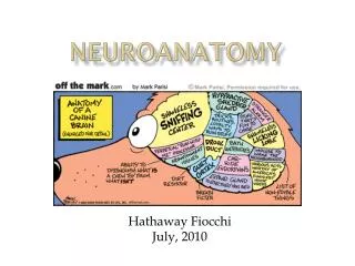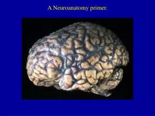
Neuroanatomy
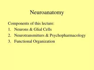
Neuroanatomy. Components of this lecture: Neurons & Glial Cells Neurotransmitters & Psychopharmacology 3. Functional Organization. Neurons, Glial Cells & Neurochemistry. Nervous System Central Nervous Peripheral Nervous System System

Neuroanatomy
E N D
Presentation Transcript
Neuroanatomy Components of this lecture: • Neurons & Glial Cells • Neurotransmitters & Psychopharmacology 3. Functional Organization
Nervous System Central Nervous Peripheral Nervous System System Brain Spinal Cord
ii. Structure of neurons 1. Cell body - contains nucleus 2. Nucleus – contains DNA 3. Dendrites – cell body (receives information) 4. Receptors – receive information using a chemical signal. 5. Axons – sends information 6. Axon hillock – junction between cell body and axon **Lowest threshold for action potential**
7. Terminal (buttons or boutons) – swelling on the surface (see slide) - Inside buttons are synaptic vesicles, packaging of neurotransmitter 8. Myelin sheath – insulation for axons - comprised of glial cells (see slide) A. In CNS it’s Oligodendrocytes B. In PNS it’s Schwann cells 9. Nodes of Ranvier – spaces between myelinating cells along the axon
11. Cell membranes cover all cells - Two layers of fat molecules - Tucked inside are channels made up of protein molecules (see slide) - Protein molecules a. Serve as receptors for NT’s – next slide b. Serve as channels for ions (Ca++, Na+, K+, and Cl-) - next slide c. Location along neuron differs in the type of channel protein d. Membranes are dynamic and alive
Two Types of Receptors (more again – soon) 1. Ionotropic Receptors fast acting ion channel/receptor complex same only a few Neurotransmitter activate them 2. Metabotropic Receptors slower acting ion channel and receptor are different most Neurotransmitters act on them
12. Cytoskeletons (Neurofilaments) inside cell provide structural support - Microfilaments - Microtubules – Fairly large, play important role in transport a. Send vesicles to the buttons where they are filled with NT. Acts like a conveyor belt.
13. Organelles within the cell a. Mitochondria – Convert glucose into energy we can use: ATP (energy source for cell) b. Endoplasmic Reticulum – Synthesis of fat molecules and protein molecules
3. Synapse - the junction between cells (neurons). - synaptic cleft - space between cells a. synapse is made of 3 parts: 1. Presynaptic cell– sending side of synapse 2. Postsynaptic – receiving side of neuron 3. Synaptic Cleft b. Purpose: promote chemical-electrical signal c. Types of Synapses: axodendritic, axosomatic axoaxonic, dendrodendritic
4. Chemical Milieu of Cellular Spaces when the neuron is “at rest” Intracellular space & extra cellular space (inside of cell membrane & outside of cell membrane) a. Cl- = Chloride (more outside than inside) b. Na+ = Sodium (more outside than inside) c. A- = Anions (more inside than outside) d. K+ = Potassium (more inside than outside)
Forces that maintain the chemical balance i. Concentration gradient – lesser concentration ii. Electrostatic pressure – attraction toward opposite charges iii. Na & K pumps – Throws out sodium and takes in potassium to keep cell balanced
5. Four states of neuronal electrical charge (potentials) a. Resting Membrane Potential -70 mV (transient state, constantly affected by forces that increase or decrease charge) b. Excitatory Post-Synaptic Potential or EPSP– Charge across the membrane becomes less negative - depolarization of the neuron (i.e. decrease negative charge from –70mV to –65mV) - Leads to an action potential
c. Inhibitory Postsynaptic Potential or IPSP Charge across the membrane becomes more negative - hyperpolarization of neurons (i.e. increase in negative charge from –70mV to –90 mV) - Reduces the likelihood of an action potential d. Action Potential or AP Charge across the membrane becomes less negative - depolarization of neurons (i.e. decrease in negative charge from –65mV to +55 mV) - charge for the AP begins at Axon Hillock - significant shift in ions
Neurotransmitters 80 plus chemical substances that provide communication between cells. Some of these are actually NTs and others are neuromodulators (i.e. they augment the activity of the NT)
Glutamate Uses both ionotropic and metabotropic receptors NT of the cerebral cortex Excitatory effect GABA Uses ionotropic receptors Most prevalent NT in the CNS Inhibitory effect Amino Acid NTs Seizures disorders are the caused by overactive Glu and/or under active GABA
Catecholamines Dopamine Norepinephrine Epinephrine Indolamines Serotonin Monoamine NTs These NTs use both reuptake and enzymes (e.g. MAO) to terminate action
Two Receptors: nicotinic receptor – uses ionotropic receptor muscarinic receptor – uses metabotropic receptor Degradation is through enzyme only: acetylcholinesterase inhibitor Acetylcholine
Neuropeptides • Long chains of amino acids • Numerous categories (see appendix VII) • One category is the ENDORPHINS • Enkephalins • Beta-endorphin
Soluble Gases • Nitric Oxide – involved in learning and memory (more on this later)
Neurotransmitters have 7 actions • Synthesized • Stored • Enzymatically destroyed if not stored • Exocytosis • Termination of release via binding with autorecptors • Binding of NT to receptors • NT is inactivated Drugs are developed that address these actions as an AGONIST (mimic the NT ) or ANTAGONIST (block the NT)
Drugs that Block Reuptake • SSRIs (Selective Serotonin Reuptake Inhibitors) • Cocaine - highly addictive, both physiologically and psychologically (see interactive CD ROM)
Tolerance & Dependence • Tolerance – state of decreased sensitivity to the drug as a result of exposure to it. functional tolerance (number of binding sites is reduced – also called “down regulation” of receptors) note: opposite phenomenon: up-regulation • Physical Dependence – caused by withdrawal symptoms (not the reason that people continue to take most drugs) • Psycholological Dependence (now called positive-incentive theory of addiction) e.g., intracranial self-stimulation studies & dopamine
Brainstem medulla, pons & midbrain a. Medulla (Myelencephalon) center for vital functions decussation of the pyramids crossing over for most nerve fibers b. Pons (Metencephalon) numerous cranial nerves reticular formation raphe nucleus and sleep
c. Midbrain (Mesencephalon) Superior Colliculus Inferior Colliculus Central Gray (periaqueductal gray) Substantia Nigra Ventral Tegmentum Schizophrenia & Parkinson’s disease
3. Cerebellum (Metencephalon) smooth coordination of practiced movements integrates sensory & motor cognitive functions (with frontal lobe)