Digital Radiography
Digital Radiography. By: Prof. Susan A. Olavidez. The Digital Radiography System. Digital radiography is performed by a system consisting of the following functional components: A digital image receptor A digital image processing unit An image management system

Digital Radiography
E N D
Presentation Transcript
Digital Radiography By: Prof. Susan A. Olavidez
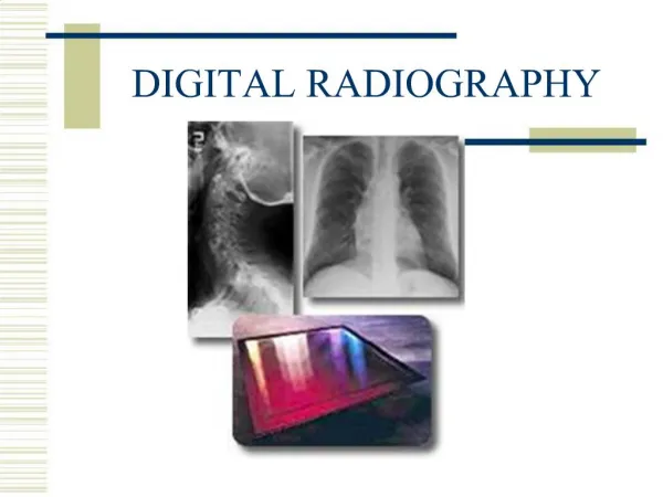
The Digital Radiography System • Digital radiography is performed by a system consisting of the following functional components: • A digital image receptor • A digital image processing unit • An image management system • Image and data storage devices • Interface to a patient information system • A communications network • A display device with viewer operated controls
The Digital Receptor • The digital receptor is the device that intercepts the x-ray beam after it has passed through the patients body and produces an image in digital form, that is, a matrix of pixels, each with a numerical value. • This replaces the cassette containing intensifying screens and film that is used in non-digital, film-screen radiography. • There are several different types of digital radiography receptors.
The Image Management System • Image management is a function performed by the computer system associated with the digital radiography process. • These functions consist of controlling the movement of the images among the other components and associating other data and information with the images. • Some of these functions might be performed by the computer component of a specific digital radiography device or by a more extensive Digital Image Management System (DIMS) that serves many imaging devices within a facility. Note: it is not unusual for the DIMS to be referred to by an older, and somewhat less appropriate name, PACS (Picture Archiving and Communications System).
Patient Information System • The Patient Information System, perhaps known as the Radiology Information System (RIS), is an adjunct to the basic digital radiography system. • Through the interface, information such as patient ID, scheduling, actual procedures performed, etc is transferred.
Image Processing • One of the major advantages of digital radiography is the ability to process the images after they are recorded. • Various forms of digital processing can be used to change the characteristics of the digital images. • For digital radiographs the ability to change and optimize the contrast is of great value. • It is also possible to use digital processing to enhance visibility of detail in some radiographs.
Digital Image Storage • Digital radiographs, and other digital medical images, are stored as digital data. • Advantages (compared to images recorded on film) include: • Rapid storage and retrieval • Less physical storage space required • Ability to copy and duplicate without loss of image quality
Communications Network • Another advantage of digital images is the ability to transfer them from one location to another very rapidly. • This can be: • Within the imaging facility to the storage and display devices • To other locations (Teleradiology) • Anywhere in the world (by means of the internet) • The total network available for transferring digital images is made up of a variety of integrated systems
Digital Image Control and Display Control • Compared to radiographs recorded and displayed on film, i.e. "softcopy", there are advantages of "softcopy" displays. • One major advantage is the ability of the viewer to adjust and optimize image characteristics such as contrast. • Other advantages include the ability to zoom, compare multiple images, and perform a variety of analytical functions while viewing the images.
The Direct Digital Radiographic Receptor • We can think of the direct digital radiographic receptor as "a digital x-ray camera".
The Direct Digital Radiographic Receptor • The receptor is in the form of a matrix of many individual pixel elements. They are based on a combination of several different technologies, but all have this common characteristic: when the pixel area is exposed by the x-ray beam (after passing through the patient's body), the x-ray photons are absorbed and the energy produces an electrical signal. This signal is a form of analog data that is then converted into a digital number and stored as one pixel in the image.
Stimualible Phosphor Radiographic Receptor • We can think of the stimualible phosphor receptor as being like a conventional radiographic intensifying screen in that it absorbs the x-ray photons and and then produces light. • The difference is that there is a delay between the x-ray exposure and the production of the light.
Stimualible Phosphor Radiographic Receptor • This is how it works: • First, a receptor (cassette) containing only a stimualible phosphor screen is exposed to record an image. At this stage the image recorded by the screen is an invisible latent image. • The next step is to process the receptor through the reader and processing unit. In this unit the screen is scanned by a very small laser beam. When the laser beam strikes a spot on the screen it causes light to be produced (the stimulation process). The light that is produced is proportional to the x-ray exposure to that specific spot. The result is that an image in the form of light is produced on the surface of the stimualible phosphor screen. • A light detector measures the light and sends the data on to produce a digitized image.
Image Formation • As the surface of the stimualible phosphor screen is scanned by the laser beam, the analog data representing the brightness of the light at each point is converted into digital values for each pixel and stored in the computer memory as a digital image.
Digital Receptor Dynamic Range • One of the significant characteristics of most digital radiographic receptors is that they have a wide dynamic range. • What that means is that the receptors respond to x-ray exposure and produce digital data over a wide range of x-ray exposure values.
Digital Radiography Quality Characteristics • Like all medical images, digital radiographs have the five specific quality characteristics as we see here • We will now see how three of these, contrast, detail, and noise are effected by the characteristics and operation of the digital system.
Digital Radiograph Contrast Characteristics • The contrast sensitivity of a digital radiographic procedure and the image contrast depend on several factors. • Two of these, the x-ray beam spectrum and the effects of scattered radiation are similar to film radiography. • What is different, and generally an advantage, with digital radiography is the ability to adjust and optimize the contrast after the image has been recorded. • This usually occurs through the digital processing of the image and then the adjustment of the window when the image is being viewed.
Digital Radiographic Detail • As in all medical images, visibility of detail is reduced and limited by the blurring that occurs at different stages of the imaging process as we see here. • What is common to both digital and film radiography are three sources of blurring: • The focal spot (depends on size and object location) • Motion (if it is present) • The receptor (generally because of light spreading within the fluorescent or phosphor screen)
Digital Radiographic Detail • What is specific to digital radiography is that additional blurring is introduced by dividing the image into pixels. Each pixel is actually a blur. As we have already observed in other modules, the size of a pixel (amount of blurring) is the ratio of the field of view (image size relative to the anatomy) and the matrix size. • Pixel size is a factor that must be considered because it limits detail in the images.
Noise in Digital Radiograph • The most predominant source of noise in digital radiography is generally the quantum noise associated with the random distribution of the x-ray photons received by the image receptor. • As we have just observed, the level of noise depends on the amount of receptor exposure used to produce an image. With digital radiography it can be adjusted over a rather wide range because of the wide dynamic range of the typical digital receptor. • The noise is controlled by using the appropriate exposure factors
REFERENCE Sprawls, Perry. The physical principles of medical imaging. Sprawls Educational Foundation Open Resource for Learning and Teaching. Retrieved: August 22, 2004, http://www.sprawls.org/resources/