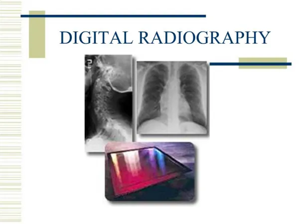
DIGITAL RADIOGRAPHY
DIGITAL RADIOGRAPHY. Digital Radiography. A “filmless” imaging system introduced in 1987 Digital radiography uses an electronic sensor, instead of “film” Requires a computerized imaging system to produce an image. No film is used, and no processing chemicals are required. Analog vs. Digital.

DIGITAL RADIOGRAPHY
E N D
Presentation Transcript
Digital Radiography • A “filmless” imaging system introduced in 1987 • Digital radiography uses an electronic sensor, instead of “film” • Requires a computerized imaging system to produce an image. • No film is used, and no processing chemicals are required.
Analog vs. Digital • “film based” • Produces a “radiograph” • Produced when x-ray photons strike the film • Shows on film as a continuous spectrum of gray shades between the extremes of white and black • The shades “flow into” one another like a painting • “sensor” is recording medium • Produces a computerized “image” • Uses an array of “pixel” elements with exact gray and discrete gray values for each pixel. • More like a mosaic pattern instead of the shades “flowing together”
Digital vs. Analog • X-ray photons • Strike sensor creating a surface electrical charge • Which is converted to digital form (digitized) • Sensor transmits digitized image to computer • Image is produced almost instantly! • Computer stores the information • X-ray photons • Interact with silver halide crystals • Produces a latent image • Chemical processing takes time • Visible image • Stored on mounts within the patient’s paper record
Analog to Digital Converter • The data acquired by the sensor is communicated to the computer in analog form, then converted by the use of the analog –to-digital-converter (ADC) • The image is displayed within seconds and may be readily manipulated to enhance diagnosis.
Types of Digital Sensors Charge-Coupled Device (CCD) Complimentary Metal Oxide Semiconductor/Active Pixel Sensor (CMOS/APS)
CCD • One of the most common digital sensors used in dental digital imaging • The CCD is a sensor that contains a silicon chip with an electric circuit built into it. • The silicon chip is sensitive to x-radiation • The electrons that compose the silicon CCD are arranged in “blocks” or picture elements known as pixels. • A pixel is the digital equivalent of the silver halide crystal on conventional film.
CCD • Unlike the silver halide crystals contained in the film emulsion, which is randomly distributed, the pixel arrangement is ordered. • The sensor will contain 307, 200 pixels! Each pixel is linked to a specific area on the computer screen. • As the x-ray photons come in to contact with the sensor, this produces an electronic charge that is connected to a specific area on the computer screen
CMOS Sensor • The CMOS differs from the CCD in the way that the pixels are read by the computer. • They claim a 25% greater resolution than CCD technology. • The CMOS sensor is also less costly to produce and the sensor has greater durability
The Computer • It stores the incoming electronic signal. • It also converts the the signal into a shade of grey that is viewed on the computer monitor. • Each pixel is represented in the computer by location and color level of the gray. • The pixel can create 256 shades of gray, but the human eye can only perceive 32 shades of gray!
Types of Digital Imaging Direct Indirect
Two Ways to Obtain a Digital Image Direct Indirect Scanning in traditional radiographs This method is inferior because the resulting image is a “copy” vs. the original • Sensor placed in mouth and exposed to x-rays. • Sensor captures image and transmits to computer monitor • Image appears within seconds
Analog vs. Digital Analog Digital Pixel – the digital equivalent of a silver halide crystal, but in an ordered arrangement 307,200 pixels on a sensor! Produce a sort of “electronic” latent image. • Silver halide crystals • Random arrangement on the film
Digital Imaging Advantages Disadvantages Initial set-up costs Image quality (on-going debate) Sensor size – thicker and less flexible for patient Legal Issues – because the image can be enhanced • Superior gray scale resolution • Decreased patient exposure to radiation • Increased speed of image viewing • Some cost reductions (no film, processing or darkroom needed) • Increased efficiency • Enhanced diagnostic image known as digital subtraction • Effective patient education tool
Other Features of Digital Imaging “Subtraction” Magnification Measurement
Digital Subtraction • With digital subtraction, the gray scale is reversed so that radiolucent images (normally black), appear white and radiopaque images (normally white), appear black. • Digital subtraction helps to eliminate distracting background information. • This feature permits the operator to remove all anatomic structures that have not changed between radiographic examinations to facilitate identification of changes in diagnostic information.