Download
1 / 45
470 likes | 867 Views
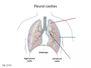
Pleural Diseases Kyphoscoliosis. MODULE E Chapters 24 & 25. Pleural Space. Visceral Pleura – attached to lungs. Parietal Pleura – attached to chest wall. Pleural space 5-10 mL of fluid secreted by the pleural cells.

E N D
Pleural DiseasesKyphoscoliosis MODULE E Chapters 24 & 25
Pleural Space • Visceral Pleura – attached to lungs. • Parietal Pleura – attached to chest wall. • Pleural space • 5-10 mL of fluid secreted by the pleural cells. • Minimizes friction as the two pleural surfaces glide over each other during inspiration and expiration.
Pleural Disease • Pleural Effusion • Accumulation of fluid in the intrapleural space. • Fluid accumulation separates the visceral and parietal pleura and compresses the lungs. • Atelectasis will develop. • Compression of heart and great vessels. • Decreased venous return. • Restrictive lung disease.
Detection of Pleural Effusions • X-ray • PA & Lateral Decubitus • Ultrasound • CT Scan
Etiology • Two Types of pleural effusions: • Transudates • Exudates
Transudates • Fluid from the pulmonary capillaries moves into the pleural space. • The fluid is thin, watery, few cells, little protein. • Clear and light straw color. • Protein content is less than 3 gm/dL. • The pleural surfaces are not involved in producing the fluid. • pH greater than 7.30.
Etiology of Transudates • Formation is the result of abnormal hydrostatic and oncotic pressures.
Etiology of Transudates • Congestive Heart Failure • Left heart failure • Hepatic Hydrothorax • Peritoneal Dialysis • Nephrotic Syndrome • Pulmonary embolism • Hypoalbuminemia
Exudates • Pleural Surfaces are diseased. • Fluid has increased protein content greater than 3 gm/dL. • Increased cellular debris . • Inflammatory process. • pH less than 7.30.
Etiology of Exudates • Malignant Pleural Effusions • Malignant mesotheliomas • Pneumonias • Tuberculosis • Fungal Diseases • Diseases of GI tract
Types of Pleural Effusions • Hydrothorax • Hydropneumothorax • Empyema • Chylothorax • Hemothorax • Loculated
Hemothorax • Blood in the pleural space. • Chest trauma • Iatrogenic hemothorax • Pulmonary embolism with infarction • Malignant disease • Also referred to as serosanguineous.
Empyema • The accumulation of pus in the pleural cavity. • Pyothorax • Develops from inflammation. • Thoracentesis will confirm the diagnosis and determine the organism.
Chylothorax • Thoracic Duct is a lymphatic channel that runs from the abdomen through the mediastinum and into the neck & empties into the left subclavian vein. • Disruption of the thoracic duct may cause leakage of chyle into the pleural space. • Malignancy, surgery and trauma.
Chylothorax • Chyle is a milky white fluid consisting mainly of fat particles.
Loculated Pleural Effusion • Confined or fixed to a single location by adhesions. • Does not move when the patient lies on his/her side.
Patient Assessment • Chest pain & decreased chest expansion • Dyspnea/WOB/Cyanosis • Cough • Shift of the PMI and trachea • Dull percussion note • Diminished BS • Tachypnea
Pulmonary Functions • Restrictive Disease • Decreased lung volumes and capacities. • Normal RV/TLC. • NO TRAPPED GAS
ABG • Small pleural effusion • Acute alveolar ventilation with hypoxemia • pH: 7.50 PaCO2: 30 torr, PaO2: 60 torr • Large pleural effusion • Acute ventilatory failure with hypoxemia • pH: 7.28 PaCO2: 55 torr, PaO2: 45 torr • Metabolic acidosis may occur if there is anaerobic metabolism ( lactic acid)
Chest X-ray Findings • Blunting of costophrenic angle. • Pleural meniscus sign. • Mediastinal shift away from affected side. • Depressed diaphragm. • A minimum of 200 – 300 mL of fluid is necessary to see a pleural effusion in an upright film. • Lateral decubitus film can pick up smaller amounts of fluid (as little as 5cc of fluid). • Atelectasis
Management of Pleural Effusions • Oxygen therapy • Thoracentesis • Chest tube • Pleurodesis • Antibiotics • Hyperinflation Protocol • Cough/deep breathing, IS, IPPB, CPAP, PEEP
Thoracentesis • Insertion of a needle into the pleural space to remove fluid or air. • Removal of a specimen for biopsy. • Therapeutically it can be used to treat a pleural effusion.
Screening for Thoracentesis • History of bleeding disorders • Platelet count • PT • Use of anticoagulants • Chest x-ray, ultrasound, CT scan
Procedure for Thoracentesis • Sign a consent form. • Administer analgesic. • Position Patient; Disinfect skin with betadine. • Assist physician with sterile mask, cap, gown and gloves. • Anesthetize the skin with 2% Lidocaine. • Insert needle until fluid level is reached.
Procedure for Thoracentesis • Withdraw 100 – 300 mL of pleural fluid with a syringe. • Withdraw needle and suture or use adhesive tape to close puncture hole. • Monitor the vital signs/PO/assess WOB. • Analyze the sample.
Color Odor RBC count WBC count Protein Glucose LDH Amylase pH Gram and AFB stains Aerobic, anaerobic, TB and fungal cultures Cytology Analysis of Pleural Fluid
Complications of Thoracentesis • Pneumothorax • Infection/empyema • Hemothorax • Subcutaneous emphysema • Air embolism • Reexpansion Pulmonary edema
Complications of Thoracentesis • Pulmonary hemorrhage. • Laceration of liver or spleen. • Pain • Mild pain for 24 hours after procedure • Shoulder pain during the procedure, indicates the tap is too low. • Needle is piercing the diaphragmatic pleura
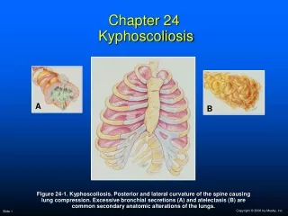
Kyphoscoliosis • Kyphosis – posterior curvature of the spine • Humpback • Scoliosis – lateral curvature of the spine • Kyphoscoliosis is a chronic disease
Anatomic Alterations • Deformity of the spine. • Compression of the lung. • Decrease lung expansion. • Atelectasis. • Hypoventilation • Inadequate cough. • Unable to mobilize secretions. • Mediastinal shift – same direction as lateral curvature.
Etiology • 10% of the US population • 1% have notable deformity • Cause unknown in 80 – 85% of cases • Idiopathic kyphoscoliosis • Pathologic conditions • Congenital vertebral defects • Vertebral disease • Neuromuscular diseases
Clinical Manifestations • Obvious thoracic deformity • Tachypnea • HR, CO, BP • Cyanosis • Weak cough with sputum production • Clubbing
Clinical Manifestations • Chest Assessment • Shift of trachea and PMI • Dull percussion note • Diminished BS/Bronchial BS • Increased tactile and vocal fremitus • Polycythemia (chronic hypoxemia/hypoxia) • Cor Pulmonale
Pulmonary Functions • Restrictive disease • Decreased volumes and capacities. • Normal flowrates. • FEV1/FVC normal.
ABG • Mild/moderate Kyphoscoliosis • Acute alveolar hyperventilation with hypoxemia • pH: 7.50 PaCO2: 30 torr, PaO2: 60 torr • Severe Kyphoscoliosis • Chronic ventilatory failure with hypoxemia • pH: 7.28 PaCO2: 55 torr, PaO2: 45 torr • Assess for CO2 retention • Watch oxygen levels
Chest X-ray • Thoracic deformity • Mediastinal shift • Radiopaque or radiodense (white) • Atelectasis • Cardiomegaly if cor pulmonale is present
Management • Oxygen Therapy • Bracing • Body brace during formative years. • Electrical stimulation • Strengthen muscles around the spine. • Surgery • Harrington and Luque Rods into the spine.
Management • Sputum C&S – antibiotics if needed • Mobilization of Bronchial Secretions • Hydration, CPT, Suctioning, IS, Bronchoscopy Deep breathing/coughing, • Hyperinflation Techniques • Cough & deep breathing, IS, IPPB, PEEP, CPAP • Mechanical Ventilation - NPV