Download
1 / 30
310 likes | 443 Views
Anatomy of a cell. Chapter 3. Typical Cell. Cells vary due to function. The typical cell exhibits the most important characteristics of many distinctive cell types. Examples of cell types: Nerve cells, muscle cells, red blood cells, gland cells, and immune cells. Functional Anatomy of Cells.

E N D
Anatomy of a cell Chapter 3
Typical Cell • Cells vary due to function. The typical cell exhibits the most important characteristics of many distinctive cell types. • Examples of cell types: Nerve cells, muscle cells, red blood cells, gland cells, and immune cells
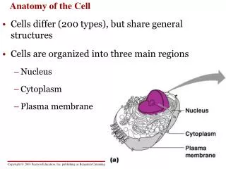
Functional Anatomy of Cells • Cell structures • Plasma membrane—separates the cell from its surrounding environment • Cytoplasm—thick gel-like substance inside of the cell composed of numerous organelles suspended in watery cytosol; each type of organelle is suited to perform particular functions • Nucleus—large membranous structure near the center of the cell
Cell Membranes • Each cell contains a variety of membranes: • Plasma membrane • Membranous organelles—sacs and canals made of the same material as the plasma membrane
Cell Membranes • Fluid mosaic model—theory explaining how cell membranes are constructed • Molecules of the cell membrane are arranged in a sheet • The mosaic of molecules is fluid; that is, the molecules are able to float around slowly • This model illustrates that the molecules of the cell membrane form a continuous sheet
Cell Membranes • Primary structure of a cell membrane is a double layer of phospholipid molecules • Heads are hydrophilic (water-loving) • Tails are hydrophobic (water-fearing) • Molecules arrange themselves in bilayers in water • Cholesterol molecules are scattered among the phospholipids to allow the membrane to function properly at body temperature • Most of the bilayer is hydrophobic; therefore water or water-soluble molecules do not pass through easily
Plasma Membranes and Proteins • A cell controls what moves through any section of membrane by means of proteins embedded in the phospholipid bilayer. • The protein acts as a gate allowing water-soluble molecules to pass through the membrane.
Cytoplasm and Organelles • Cytoplasm—gel-like internal substance of cells that includes many organelles suspended in watery intracellular fluid called cytosol
Cytoplasm and Organelles • Two major groups of organelles: • Membranous organelles are specialized sacs or canals made of cell membranes • Nonmembranous organelles are made of microscopic filaments or other nonmembranous materials
Organelles • Endoplasmic reticulum (Highway system of the cell) • Made of canals with membranous walls and flat, curving sacs arranged in parallel rows throughout the cytoplasm; extend from the plasma membrane to the nucleus • Proteins move through the canals
ER • Two types of ER: 1) Rough ER: Ribosomes on the outer surface of the organelle 2) Smooth ER: Synthesizes certain lipids and carbohydrates and creates membranes for use throughout cell
nonmembranous structure • Ribosomes in the endoplasmic reticulum make proteins for “export” or to be embedded in the plasma membrane; free ribosomes make proteins for the cell’s domestic use
Golgi apparatus • Golgi apparatus (UPS of cell): flat-like pancake organelle that packages and delivers proteins and other organic molecules throughout the cell.
Lysosomes • Lysosomes (miniature stomach)- enzymes in lysosomes digest the protein structures of defective cell parts, including plasma membrane proteins, and particles that have become trapped in the cell
Mitochondria • Mitochondria (power plant of cell)- mitochondrial enzymes catalyze series of oxidation reactions that provide about 95% of cell’s energy supply • Each mitochondrion has a DNA molecule, allowing it to produce its own enzymes and replicate copies of itself
Nucleus • Consists of nuclear envelope (composed of two membranes each with essentially the same molecular structure as plasma membrane) surrounding nucleoplasm; nuclear envelope has holes called nuclear pores
Cytoskeleton • The cell’s internal supporting framework made up of rigid, rodlike pieces that provide support and allow movement and mechanisms that can move the cell or its parts (Figure 3-13)
Centrosome • An area of the cytoplasm near the nucleus that coordinates the building and breaking of microtubules in the cell- Important in cell division