Download

1 / 97
970 likes | 1.02k Views
Learn about primary & secondary lymphoid organs, bone marrow, thymus, lymph nodes, spleen, MALT, & histological structures of thymus.

E N D
LYMPHOID TISSUE This resource is licensed under the Creative Commons Attribution Non-Commercial & No Derivative Works License
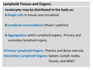
Objectives Appreciate that the lymphoreticular system is divided into primary and secondary lymphoid organs. Recognise that the structure of the BONE MARROW and THYMUS provides an ideal environment for B cell and T cell differentiation. Describe how the structure of the LYMPH NODE is well adapted for filtering antigens from the tissue fluid and initiating immune responses. Describe the structure of the SPLEEN and appreciate that this is related to its role in initiating immune responses to antigens in the blood. List the important components of the MUCOSAL ASSOCIATEDLYMPHOID TISSUES (MALT) and describe how their structure relates to their function in protecting mucosal surfaces.
Bone MarrowSee Blood and Haemopoiesis (year 1 of course 004)
SLIDE 147 Thymus (cat) Examine the thymus at very low magnification. Identify; a. Capsule. b. Thymic lobules with cortical and medullary zones. 1.0 mm
SLIDE 147 Thymus (cat) c c : capsule m : medulla m septum Examine the thymus at very low magnification. Identify; a. Capsule. b. Thymic lobules with cortical and medullary zones. cortex m m thymic lobule 1.0 mm
SLIDE 147 Thymus (cat) Examine the thymus at low magnification. Identify; a. Capsule. b. Thymic lobules with cortical and medullary zones. 0.5 mm
SLIDE 147 Thymus (cat) Examine the thymus at low magnification. Identify; a. Capsule. b. Thymic lobules with cortical and medullary zones. cortex medulla capsule of connective tissue continues as septa between the lobules 0.5 mm
SLIDE 147 Thymus (cat) What are the major differences in the histological structure of the thymus in the cortical and medullary zones? 100 µm
What are the major differences in the histological structure of the thymus in the cortical and medullary zones? Cortex: tightly packed with small lymphocytes, few stromal cells visible. SLIDE 147 Thymus (cat) What are the major differences in the histological structure of the thymus in the cortical and medullary zones? cortex : packed with dark staining lymphocytes 100 µm
What are the major differences in the histological structure of the thymus in the cortical and medullary zones? Cortex: tightly packed with small lymphocytes, few stromal cells visible. Medulla: fewer lymphocytes, more non-lymphoid cells visible (e.g. Epithelioreticular cells, macrophages) and more obvious blood vessels. SLIDE 147 Thymus (cat) medulla : fewer lymphocytes What are the major differences in the histological structure of the thymus in the cortical and medullary zones? cortex : packed with dark staining lymphocytes 100 µm
SLIDE 147 Thymus (cat) In which part of the thymic lobule would you expect to see mature lymphocytes and can immature and mature lymphocytes be distinguished morphologically? 50 µm
In which part of the thymic lobule would you expect to see mature lymphocytes and can immature and mature lymphocytes be distinguished morphologically? Immature in cortex. Mature in medulla. SLIDE 147 Thymus (cat) medulla of thymus lymphocytes (dark staining) In which part of the thymic lobule would you expect to see mature lymphocytes and can immature and mature lymphocytes be distinguished morphologically? pale staining non-lymphoid cells (epithelioreticular cells, macrophages) H BV BV : blood vessel H : Hassall’s corpuscle 50 µm
In which part of the thymic lobule would you expect to see mature lymphocytes and can immature and mature lymphocytes be distinguished morphologically? Immature in cortex. Mature in medulla. It is difficult to distinguish between the two types of cell on morphology but you could look for expression of cell surface markers. e.g. CD4 & CD8 : double negative→double positive→single positive. SLIDE 147 Thymus (cat) medulla of thymus lymphocytes (dark staining) In which part of the thymic lobule would you expect to see mature lymphocytes and can immature and mature lymphocytes be distinguished morphologically? pale staining non-lymphoid cells (epithelioreticular cells, macrophages) H BV BV : blood vessel H : Hassall’s corpuscle 50 µm
SLIDE 147 Thymus (cat) medulla cortex What is the function of epithelioreticular cells, how can they be identified in this section? 25 µm
What is the function of epithelioreticular cells, how can they be identified in this section? They provide the structural framework of the thymus and secrete growth factors as well as essential cell-cell interactions with lymphocytes. These cells are bigger, lighter staining and more heterogeneous in shape compared to the lymphocytes. SLIDE 147 Thymus (cat) medulla cortex E E lymphocytes E What is the function of epithelioreticular cells, how can they be identified in this section? Hassall’s corpuscle E E 25 µm E : epithelioreticular cells
SLIDE 147 Thymus (cat) What is the distinguishing feature of Hassall’s corpuscles and are they found anywhere other than in thymic medulla? 25 µm
What is the distinguishing feature of Hassall’s corpuscles and are they found anywhere other than in thymic medulla? They look like a section through an onion (degenerate epithelial cells forming squames). These are only found in the thymus. SLIDE 147 Thymus (cat) 25 µm concentric whorls of degenerating epithelioreticular cells forming Hassall’s corpuscles (thymic corpuscles).
SLIDE 143 Lymph node (cat) Identify : a. cortex (outer layer). d. germinal centre. b. paracortex. e. fibrous capsule. c. medulla (deeper layer). 1.0 mm
SLIDE 143 Lymph node (cat) C : cortex PC : paracortex M : medulla G : germinal centre H : hilum fibrous capsule M H Identify : a. cortex (outer layer). d. germinal centre. b. paracortex. e. fibrous capsule. c. medulla (deeper layer). PC G C 1.0 mm
SLIDE 143 Lymph node (cat) The sinuses characterise the structure of the lymph node. Try to visualise how the lymph carrying the antigen enters the subcapsular sinus and flows through the lymph node via cortical and medullary sinuses. 1.0 mm
SLIDE 143 Lymph node (cat) lymph reaches subcapsular sinus from afferent lymphatics. The sinuses characterise the structure of the lymph node. Try to visualise how the lymph carrying the antigen enters the subcapsular sinus and flows through the lymph node via cortical and medullary sinuses. 1.0 mm
SLIDE 143 Lymph node (cat) lymph reaches subcapsular sinus from afferent lymphatics. flows through cortical sinuses in trabeculae. The sinuses characterise the structure of the lymph node. Try to visualise how the lymph carrying the antigen enters the subcapsular sinus and flows through the lymph node via cortical and medullary sinuses. 1.0 mm
SLIDE 143 Lymph node (cat) lymph reaches subcapsular sinus from afferent lymphatics. flows through cortical sinuses in trabeculae. flows through medullary lymph sinuses. The sinuses characterise the structure of the lymph node. Try to visualise how the lymph carrying the antigen enters the subcapsular sinus and flows through the lymph node via cortical and medullary sinuses. 1.0 mm
SLIDE 143 Lymph node (cat) lymph reaches subcapsular sinus from afferent lymphatics. flows through cortical sinuses in trabeculae. flows through medullary lymph sinuses. exits from hilus of lymph node via efferent lymphatics. The sinuses characterise the structure of the lymph node. Try to visualise how the lymph carrying the antigen enters the subcapsular sinus and flows through the lymph node via cortical and medullary sinuses. 1.0 mm
SLIDE 143 Lymph node (cat) lymph reaches subcapsular sinus from afferent lymphatics. flows through cortical sinuses in trabeculae. flows through medullary lymph sinuses. exits from hilus of lymph node via efferent lymphatics. The sinuses characterise the structure of the lymph node. Try to visualise how the lymph carrying the antigen enters the subcapsular sinus and flows through the lymph node via cortical and medullary sinuses. 1.0 mm
SLIDE 143 Lymph node (cat) Identify : a. cortex (outer layer). e. medullary sinuses. b. paracortex. f. fibrous capsule. c. medulla (deeper layer). g. connective tissue trabeculae. d. medullary cords. 0.5 mm
SLIDE 143 Lymph node (cat) Ms : medullary sinuses Mc : medullary cords capsule Identify : a. cortex (outer layer). e. medullary sinuses. b. paracortex. f. fibrous capsule. c. medulla (deeper layer). g. connective tissue trabeculae. d. medullary cords. Mc Mc Ms trabeculae medulla cortex Ms paracortex 0.5 mm
SLIDE 143 Lymph node (cat) Identify : a. cortex. d. sub-capsular sinus. b. fibrous capsule. c. connective tissue trabeculae. 100 µm
SLIDE 143 Lymph node (cat) sub-capsular sinus cortex capsule Identify : a. cortex. d. sub-capsular sinus. b. fibrous capsule. c. connective tissue trabeculae. trabecula cortex 100 µm
SLIDE 143 Lymph node (cat) Part of the trabecula of a lymph node. The sub-capsular sinus continues as cortical sinuses in the trabeculae. Smooth muscle fibres can be seen in the trabeculae. 50 µm
SLIDE 143 Lymph node (cat) cortex Part of the trabecula of a lymph node. The sub-capsular sinus continues as cortical sinuses in the trabeculae. Smooth muscle fibres can be seen in the trabeculae. trabeculae with smooth muscle fibres cortical sinus cortex 50 µm
SLIDE 143 Lymph node (cat) Under higher magnification distinguish different cell types in the germinal centre and peripheral corona of follicles and diffuse paracortex. 100 µm
SLIDE 143 Lymph node (cat) paracortex trabecula capsule Under higher magnification distinguish different cell types in the germinal centre and peripheral corona of follicles and diffuse paracortex. peripheral corona germinal centre 100 µm
SLIDE 143 Lymph node (cat) paracortex peripheral corona germinal centre Under higher magnification distinguish different cell types in the germinal centre and peripheral corona of follicles and diffuse paracortex. 50 µm
SLIDE 143 Lymph node (cat) What are the major cell types found in the paracortex and how does this differ from the cells present in the follicles or medulla? 50 µm
What are the major cell types found in the paracortex and how does this differ from the cells present in the follicles or medulla? T cells, interdigitating DCs and macrophages in paracortex. B cells and follicular DCs in follicles. Macrophages and plasma cells in medulla. SLIDE 143 Lymph node (cat) 50 µm
SLIDE 143 Lymph node (cat) Examine the medulla with medullary cords and sinuses. 250 µm
SLIDE 143 Lymph node (cat) capsule cortex paracortex Examine the medulla with medullary cords and sinuses. Medulla : MC : medullary cords MS : medullary sinuses MC blood vessels MS 250 µm
SLIDE 143 Lymph node (cat) The medulla with medullary cords and sinuses lined with an endothelium containing lymphocytes, plasma cells and macrophages. 100 µm
SLIDE 143 Lymph node (cat) medullary cords The medulla with medullary cords and sinuses lined with an endothelium containing lymphocytes, plasma cells and macrophages. medullary sinuses 100 µm
SLIDE 125 Lymph node (dog) View the larger of the two sections. This has been stained to demonstrate reticular fibres. 2.0 mm
SLIDE 125 Lymph node (dog) Identify the main regions of this lymph node stained for reticular fibres. 1.0 mm
SLIDE 125 Lymph node (dog) trabeculae capsule cortex Identify the main regions of this lymph node stained for reticular fibres. paracortex medulla 1.0 mm
SLIDE 125 Lymph node (dog) At high magnification the network of reticular fibres can be seen in this area of medulla. What is this protein fibre composed of? 25 µm
SLIDE 125 Lymph node (dog) medullary sinus At high magnification the network of reticular fibres can be seen in this area of medulla. What is this protein fibre composed of? Collagen type III. medullary cords R R R : reticular fibres 25 µm
SLIDE 145 Spleen (rat) Whole section seen under low magnification. Identify : a). capsule. d). central artery. b). white pulp. c). red pulp. 1.0 mm
SLIDE 145 Spleen (rat) R Whole section seen under low magnification. Identify : a). capsule. d). central artery. b). white pulp. c). red pulp. R W W W central artery capsule R W : white pulp R : red pulp 1.0 mm
SLIDE 145 Spleen (rat) R What features distinguish the spleen from lymph nodes and other lymphoid tissues? R W W W central artery capsule R W : white pulp R : red pulp 1.0 mm
SLIDE 145 Spleen (rat) R What features distinguish the spleen from lymph nodes and other lymphoid tissues? Central arteries in the white pulp. No cortex and medulla. R W W W central artery capsule R W : white pulp R : red pulp 1.0 mm
Spleen – Dog ▪ Cadaver in right lateral recumbency. Abdominal wall has been removed. Ribcage & diaphragm intact. Left ribcage is retracted laterally with forceps. ▪ Identify : tendinous & muscular portions of diaphragm left, lateral & left medial lobes of liver right medial lobe of liver fundus & body of stomach greater curvature of stomach & gastrosplenic ligament spleen