Primary Open Angle Glaucoma: Pathophysiology and Management
620 likes | 2.98k Views
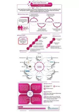
Learn about primary open angle glaucoma, its risk factors, clinical features, and management. Explore the etiopathogenesis, pathophysiology, and aqueous humor formation in this eye disease.

Primary Open Angle Glaucoma: Pathophysiology and Management
E N D
Presentation Transcript
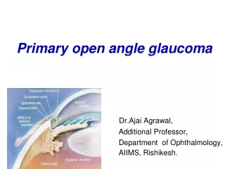
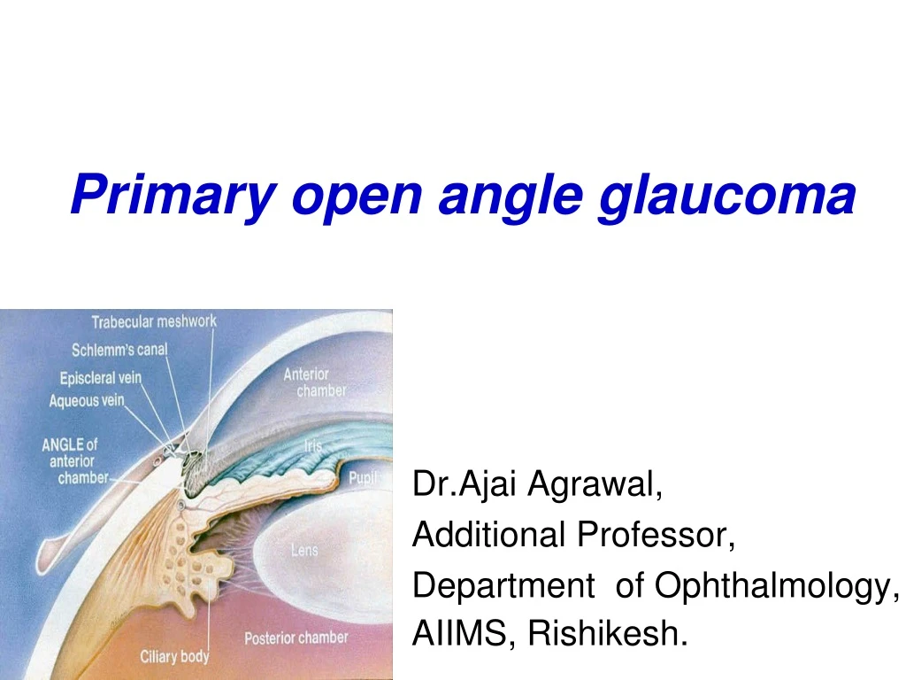
Primary open angle glaucoma Dr.Ajai Agrawal, Additional Professor, Department of Ophthalmology, AIIMS, Rishikesh.
Acknowledgement • Kanski’s Clinical Ophthalmology (8th Edition). • Becker- Schaffer’s Diagnosis and therapy of The Glaucomas (8th Edition). • Comprehensive Ophthalmology (A.K.Khurana) (7th Edition).
Learning Objectives • At the end of this class the students shall be able to : • Define primary open angle glaucoma(POAG). • Comprehend the pathophysiology and risk factors of POAG. • Understand the clinical features of POAG. • Understand the fundamentals of managing primary open angle glaucoma
Question Glaucoma is defined as: • a. a group of diseases that have in common a characteristic optic neuropathy associated with increased intraocular pressure. • b. a group of diseases that have in common a characteristic optic neuropathy with associated visual function loss. • c. a group of diseases that have in common high intraocular pressure with or without optic neuropathy. • d. a group of diseases that have in common a characteristic optic neuropathy with poor visual acuity.
Definition of POAG • Chronic, progressive optic neuropathy characterised by morphological changes at the optic disc and retinal nerve fibre layer leading to characteristic visual field changes, in the absence of other ocular diseases or congenital anomalies (with or without a raised IOP).
Etiopathogenesis • Multifactorial aetiology • Risk factors include: • Elevated Intra Ocular Pressure(IOP) (More than 21 mm Hg) • Optic disc cupping • Increasing Age : More common in 5th to 7th decades • Race: More common and severe in Black population
Etiopathogenesis • Heredity/ Family History: Risk of about 10% in siblings; 4% in off springs • Diabetes • Systemic Hypertension • Myopia • Thin central corneas • Steroid usage • ??Migraine, Cigarette smoking
Pathophysiology of POAG • Decrease in aqueous outflow facility due to increased resistance to outflow leads to rise in IOP • Two theories of axonal loss in optic disc • 1. Mechanical: Distortion of lamina cribrosa leading to impaired axoplasmic flow 2. Vascular: Optic disc ischaemia with defective autoregulation of blood vessels
FORMATION OF AQUEOUS HUMOR CILIARY PROCESSES -approx. 70-80 radial folds in the pars plicata which form the site of aqueous production. -Zonular fibers attach primarily in the valleys of the ciliary processes and also along the pars plana
FORMATION PROCESSES DIFFUSION SECRETION (80-90%) ULTRA- FILTRATION
Formation of aqueous humor • Diffusion and ultrafiltration are both passive mechanisms so no active cellular participation occurs. • Active secretion is an active process. • Rate of formation of aqueous humor in a healthy human eye is- 2 - 3 microlitres/minute
-Marked deficit of proteins -Marked excess of Ascorbate -Excess of Lactate -Excess of Chloride & certain amino acids AQUEOUS PLASMA 0.024 7.0 gm/dl gm/dl 1.06 0.04 micromol/ml micromol/ml 4.5 1.9 micromol/ml micromol/ml Differences between aqueous humor & plasma
Functions of aqueous humor *Maintaining IOP : -important for early ocular development & maintaining global integrity throughout life. *Serves as a vascular system for the avascular structures of the eye: cornea, lens & TM. - by providing substrates & nutrients & removing metabolites.
Functions of aqueous humor *Delivering high concentration of Ascorbate: - scavenges free radicals & protects against UV rays & other radiations. *Local paracrine signaling & immune responses. *Colourless & transparent medium as part of eye’s optical system.
Major amount of aqueous humor leaves the eye by BULK FLUID FLOW i.e. fluid flows along normal pressure gradient through non-energy dependent process
Ciliary processes ↓ Aqueous Humor in PC ↓ through pupil ↓ Anterior Chamber
Trabeculo-canalicular outflow *It is the main outlet for aqueous from the AC *70-90% of total aqueous is drained by this route
TRABECULAR MESHWORK -A sponge work of connective tissue beams arranged as super-imposed perforated sheets. - Extracellular spaces contain hydrophilic glycosaminoglycans & collagen.
JUXTACANALICULAR (ENDOTHELIAL) MESHWORK - Outermost portion of TM which mainly offers the normal resistance to aqueous outflow - Connects the corneoscleral meshwork with schlemm’s canal
Veins from the anterior part of ciliary body form the Ciliary venous plexus Anterior ciliary veins & Episcleral veins communicate with Schlemm’s canal
Schlemm’s Canal 20-30 Collector channels Aqueous Vein Intra-scleral venous plexus Episcleral venous plexus & Anterior Ciliary vein
UNCONVENTIONAL OUTFLOW *responsible for 10-25% of total aqueous outflow
Trans-corneal outflow • Aqueous humor from anterior chamber goes into tear film through cornea. • Very little aqueous passes through this pathway. • Total volume of fluid transferred is limited by high hydraulic resistance of the cornea.
Clinical features of POAG Symptoms • Usually asymptomatic in early cases • Mild headache and eye ache • Frequent changes in presbyopic glasses • Delayed dark adaptation • Loss of peripheral vision • Loss of central vision(late cases)
Signs of POAG • Normal anterior segment • Pupil reaction to light may be sluggish(in advanced cases only) • Elevated IOP(More than 21 mm Hg) with diurnal variation more than 5-8 mmHg • Opticdisc changes (Progressive, asymmetric) • Visual field defects
Optic disc changes in glaucoma • Early changes • Retinal nerve fibre layer atrophy • Vertically oval cup • Asymmetry of the cups(More than 0.2 difference) • Large cup(CD more than 0.6) • Splinter haemorrhages
Advanced glaucomatous disc changes • Marked cupping (More than 0.7) • Thinning of NRR (Neuroretinal rim) • Lamellar dot sign • Vascular alterations • Nasal shifting of retinal vessels • Bayonetting sign(convoluted path due to NRR loss) • Baring of circumlinear vessels and overpass vessels • Glaucomatous optic atrophy
Normal Optic Disc Glaucomatous optic disc
Glaucomatous optic disc Is this a normal or glaucomatous disc ?
Recording and documenting disc changes • Serial drawings (10 square grid) after seeing fundus by ophthalmoscopy/slit lamp with +90D/+78D lens • Disc photography • HRT(Heidelberg retinal tomography) • OCT (Optical coherence tomography) • NFA(Nerve fibre analyser)
Visual field defects in glaucoma • Arcuate nerve fibres in the superior and inferior temporal portions of the optic disc: Most sensitive to damage • Macular fibres : Most resistant to damage CENTRAL VISION IS PRESERVED TILL THE LAST IN GLAUCOMA
Progression of field defects • Isopter contraction: Generalised field constriction • Baring of blind spot : Non specific (Exclusion of blind spot from central field) • Paracentralscotoma: Wing shaped and occurs above or below the blind spot in the Bjerrum’s area(10-25 degrees from fixation) Is the earliest clinically significant defect
Progression of field defects • Seidel’s scotoma: sickle shaped Due to joining of blind spot and paracentralscotoma • Bjerrum’s/Arcuatescotoma: Extension of Seidel’s scotoma to reach the horizontal line. • Double arcuate/ring scotoma
Progression of field defects • Roenne’s central nasal step: Sharp right angled defect at the horizontal meridian when arcuatescotomas run in different arcs • Peripheral field defects • Advanced defects Residual Tubular vision Temporal island of vision
Quantification of visual field defects • Visual field analyzer Kinetic perimeter Static perimeter (automated) Testing more than once is required before final interpretation
Double arcuate 10-2- Advanced VFD , macular split
Diagnostic work up/Investigations • Tonometry • Goniscopy: Open angles • Perimetry: To detect visual field defects • Slit lamp examination: To rule out causes of secondary open angle glaucoma • Fundus examination to document optic disc changes • Diurnal variation testing • Provocative testing: Water drinking test