PRIMARY OPEN ANGLE GLAUCOMA(POAG)
PRIMARY OPEN ANGLE GLAUCOMA(POAG). BY: Katidjah Rahim Norsuriati Abd Khamis Siti Zawani Mistoh @ Mastan. DEFINITION.

PRIMARY OPEN ANGLE GLAUCOMA(POAG)
E N D
Presentation Transcript
PRIMARY OPEN ANGLE GLAUCOMA(POAG) BY: KatidjahRahim NorsuriatiAbdKhamis SitiZawaniMistoh @ Mastan
DEFINITION • Primary open-angle glaucoma is described as bilateral, non-congestive increase of IOP in absence of angle closure leading to optic nerve damage from multiple possible causes that is chronic and progresses over time, with a loss of optic nerve fibers.
INCIDENCE • Occur equally between male and female • Occur above age of 40 years old • More in black people (African – American) • Bilateral but one eye preceded before the other
RISK FACTORS • AGE: Increasing risk after age 40 and continues to increase with each additional decade. Aging also can cause drainage channels in the trabecular meshwork to shrink or narrow, which slows the outflow of fluid from the eye.
CERTAIN MEDICAL PROBLEMS: • Diabetes • High myopia • The use of oral or inhaled steroids • Migraine headaches, • high blood pressure, narrowed blood vessels (vasospasm) and cardiovascular disease.
EYE ABNORMALITIES • Pseudoexfoliationsyndrome causes proteins in the eye's natural lens, iris and other structures to slough off and clog the eye's drainage system. • RACE: three to four times more common in African-Americans than in whites. • FAMILY HISTORY:threeto four times higher if one or more of your parents and siblings have the disease.
CLINICAL PICTURE • SYMPTOMS - no acute symptom, and it may pass unnoticed until complete loss of vision. The symptoms may be: a) headache or feeling of fullness b) delayed dark adaptation c) early presbyopiadue to pressure on the ciliary nerves and weakness of ciliary muscle d) blurring of vision and field loss are late symptoms
1-TENSION: (normal IOP is 10:20 mmHg by applanationtonometer) • Applanationtonometryshould be used to avoid the factor of sclera rigidity • High IOP in presence of open angle is diagnostic but normal tension does not exclude POAG because early stages of the disease show wide fluctuation of the IOP , in this case we must resort to one of the following methods:
(A)IOP IN BOTH EYES: -Normally does not exceed 4 mmHg, 8 mmHg or more are diagnostic. (B)DIURNAL VARIATION • normally IOP is highest in the morning and goes to the minimum in the late evening but the variation is never more than 4 mmHg • the patient is hospitalized and IOP is measured every 4 hours for 24 hours . if diurnal variation exceed 8 mmHg, POAG is diagnosed.
(C)PROVOCATIVE TEST: • the aim of these test is to increase aqueos formation with faulty in the drainage systemrise of IOP 1- water drinking test: to measure the rise in IOP after drinking one liter of water. The IOP is measured every 15 minutes for 1-2 hours. A rise of 8mmHg or more is diagnostic. 2- Priscol test: 10mg of priscol is injected sub conjunctively. A rise of 11-13 mmHg is Suspicious and 14 mmHg is pathological. (D)TONOGRAPHY
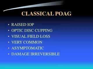
2-OPTIC NERVE HEAD CHANGES (GLAUCOMATOUS CUPPING) • The normal disc is pink in colour and 1.5mm in diameter. It is divided into a central pale depression called optic cup (normally 0.3 of the disc in diameter) and a neuro-retinal rim sorrunding it. • The rim is composed of: • Papillo-macular bundle: from the macula (temporally) • Superior arcuate bundle: from superior temporal retina (up) • Inferior arcuate bundle: from inferior temporal retina (down) • Nasal bundle: from nasal retina (nasally) *the arcuate bundles are susceptible to early damage in glaucoma producing vertical enlargement or notching of the cup and early central field changes.
Causes of glaucomatous cupping • mechanical factor: the rise of IOP lead to bowing of lamina cribrosa backward (weak area) • ischemic optic neuropathy: sclerosis of small optic nerve vesselsdegeneration of optic N.F small empty spaces which coalescecavernousdegenerationCTcontractionbackward retraction of the laminaglaucomatous cupping.
Methods to record optic disc shape • Fundus photography • Nerve fiber layer analyzer • Conofocal laser opthalmoscope (CLO) • Optical coherence tomography (OCT)
Characteristic of glaucomatous optic disc cupping: 1-vertically oval 2- pale disc 3- wide cup 4- deep cup 5- Asymmetric cupping 6- Undermined edges (interrupted blood vessel at the cup margin) 7- Nasal shift of blood vessel 8- Splinter hemorrhage (flame shaped at the cup edge) 9- Progression of cupping (most important sign)
3-VISUAL FIELD CHANGES: • Central field changes: -The central field (30 degrees) is examined by Bjerrum screen (campimetry) or the recent automated perimetry.
-Central field changes include: (a)Isolated paracentralscotoma: • they are found early in glaucoma within Bjerrum area (central 20 degrees)
(b)Baring of the blind spot: - exclusion of the blind spot from central field (c) Seidl’sscotoma: - extension of the blind spot above or below in a sickle shape manner.
(d) Arcuate or Bjerrumscotoma: - an arcuatescotoma continuous with the blind spot - concentric with point of fixation and ends in the horizontal meridian - it follows the arcuate fibers (typical nerve fiber bundle defect)
(e) Ring or annular scotoma - fusion of upper and lower arcuatescotoma.
Peripheral field changes • Generalized contraction: more on the nasal side • Ronnie nasal step: the nasal contaction extend to the horizontal raphe • Terminal field: tubular field (central 5-10 degrees) with temporal island
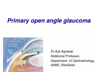
GONIOSCOPICALLY OPEN ANGLE • Gonioscopyto visualise the iridocorneal angle utilises a contact lens to avoid the problem of total internal reflection which normally makes all angle structures invisible. ( The large difference in refractive index between the cornea and air has to be minimised. )
The Goldmanngonioscopehas a highly curved anterior surface which needs to be filled with about 3 drops of normal saline or hypromellose before application to the anaesthetised cornea.
Under the Shaffer angle grading system each quadrant is given a grade from:- • 0 :is closed (either contact or adhesion) • I :10-15 degrees • II :15 to 25 degrees • III :25 to 35 degrees • IV :40 or more derees
TREATMENT • The options for treatment of glaucoma include one or more of the following: 1. Medication 2. Laser trabeculoplasty 3. Filtration and other surgery
Surgical treatment Indication : • Medical and laser treatment fail • Visual field deteriorates • Poor patient compliance • Inadequate ophthalmic care
Aim Of Surgery Is to create a new pathway for aqueous to flow from A.C through a scleral opening into the subconjunctival or sub- Tenon’s space.
Operation For POAG A)External Fistulizing Procedure • Subscleraltrabeculectomy • Trabeculectomy plus mitomycin C (intra-operative) or 5FU (post-operative) • Laser sclerotomy • Non-penetrating fistulizing procedure e.gVisco-canaloplasty
B) Seton (Tube-Shunt) Surgery C) Cilliary Body Destructive Surgeries • Cyclocyotherapy • Cyclodiathermy • Cyclophotocoagulation • Trans scleral (YAG OR diode laser) • Trans pupillary by Argon laser
D) Cyclodialysis (Internal Fistulizing Procedure) *Indication : • Aphakic glaucoma • Posterior lens dislocation • Congenital aniridia