Download
1 / 17
170 likes | 440 Views
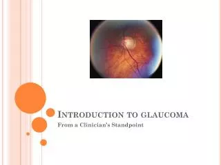
Glaucoma & primary disorders of the endothelium. Jorge L. Fernandez Bahamonde, MD. Definition. Primary corneal endothelium abnormalities directly associated with the changes causing glaucoma. ICE syndrome. Essential iris atrophy (Progressive iris atrophy). Iris-nevus syndrome (Cogan-Reese).

E N D
Glaucoma & primary disorders of the endothelium. Jorge L. Fernandez Bahamonde, MD.
Definition. • Primary corneal endothelium abnormalities directly associated with the changes causing glaucoma. • ICE syndrome. • Essential iris atrophy (Progressive iris atrophy). • Iris-nevus syndrome (Cogan-Reese). • Chandler’s syndrome. • Posterior polymorphous dystrophy.
Progressive iris atrophy. • Described by Harms in 1903. • Secondary glaucoma + iris holes. • Characteristics. • Prominent iris atrophy. • Iris stromal and/or full thickness holes. • Strech & melting holes. • Early PAS with central & circumferential progression. • Pupil distortion, ectropion uvea
Iris-nevus (Cogan-Reese). • 1969, initial two cases ended in enucleation. • Iris pigmented lesions. • Multiple. • Pedunculated or nodular. • Diffuse and velvety changes. • Darker iris color.
Iris-nevus (Cogan-Reese). • Ectropion uvea, ectopic pupil, breaks in stroma. • PAS, corneal edema, secondary glaucoma.
Chandler’s • 1956, somewhat similar to iris atrophy but corneal changes more prominent. • Iris changes. • Slight correctopia and mild stromal atrophy. • Corneal edema even with “normal” IOP. • Endothelium: hammered silver appearance. • Secondary glaucoma. • Progressive angle closure by PAS.
Pathophysiology of ICE • Endothelialization of the anterior chamber. • Abnormal corneal endothelium spreads through TM and iris. • Glaucoma more advanced than PAS suggest.
Epidemiology & Etiology ICE. • Young to middle aged woman. • Male:female 1:2 to 1:5. • Typical patient white without family hx. • Unilateral, no genetic predisposition. • Theories. • Inflammatory, low grade flare? • Vascular, insufficiency of iris vessels. • Primary Iris Defect. • Membrane theory. • Abnormal corneal endothelium invade A/C.
Etiology ICE, cont. • Viral endotheliitis 10-20 year old. • HSV, Epstein Barr? • Conversion of corneal endothelium into ICE cells. • Migratory behavior toward the iris. • Aberrant BM. • Past herpes keratouveitis. • HSV DNA localized only in endothelium.
Specular Microscopy & ICE. • ICE cells. • Larger and more pleomorphic than regular endothelial cells. • In some cases replace all corneal endothelium.
DD of ICE. • Fuch’s endothelial dystrophy. • PPD. • CHED. • Post-surgery. • Anterior segment dysgenesis. • HSV. • Iris nevus. • JXG.
Posterior Polymorphous Dystrophy. • Deep corneal lesions of varied shapes, easily seen with retroillumination. • Nodular. • Vesicular, blister like. • Gray halos. • Gray-white opacities. • Thickening of Descemet. • Bands, confused with tears. • Abnormal pleomorphic endothelium.
Posterior Polymorphous Dystrophy. Epidemiology. • A. Dominant. • Some recessives cases. • Bilateral. • Asymmetric, rarely unilateral. • Age onset. • Difficult, most patient are asymp. • Mostly non-progressive. • Some develop corneal decompensation. • Congenital cases: corneal edema.
Posterior Polymorphous Dystrophy. Associations. • Anterior segment dysgenesis. • From prominent Schwalbe’s to ectropion uvea. • Elevated IOP in 15% cases of PPD. • Small % associated to Alport’s syndrome. • Linkage to 20 q in one family.
Glaucoma and ICE. • Common, secondary angel closure. • Closure of the angle by coverage of abnormal endothelium and BM, followed by PAS. • Difficult to manage. • ICE in the DD of unilateral secondary angle closure in young population.
Rx ICE. • Aq. Suppressants. • ALT/SLT contraindicated. • Filtering surgery. • Late failure due to proliferation of endothelial membrane into filtering bleb. • MMC. • Lots of PF. • Hypertonic saline. • PKP.