Download

1 / 17
170 likes | 189 Views
Explore the different chambers and valves of the heart, as well as the characteristics of cardiac muscle cells. Discover the role of autorhythmic cells and the excitation-contraction coupling process.

E N D
Cardiac Physiology • The heart: chambers, the valves • Cardiac muscle cells • Some cardiac muscle cells are autorhythmic • Arrangement of cardiac muscle cells • Excitation-contraction coupling
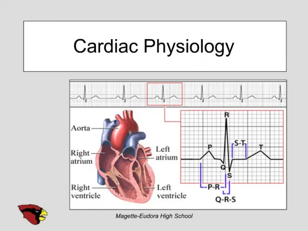
Atrioventricular (AV) Valves • guard the passageway between the atria and the ventricles • Tricuspid valve between right atrium and right ventricle • Bicuspid (mitral) valve between left atrium and left ventricle
Semilunar valves • Between ventricles and arteries • Pulmonary valve between right ventricle and pulmonary artery • Aortic valve between left ventricle and aorta
All myocardial cells • Gap junctions at intercalated discs, waves of depolarization spread from one cell to another
Autorhythmic myocardial cells • (pacemakers) are small myocardial cells with few contractile fibers • Spontaneously generate action potentials • Enables the heart to contract without any outside signal • The heart is myogenic: signal for contraction originates from heart muscle itself
Most myocardial cells • Remaining myocardial cells are striated • Have sarcomeres • Much smaller than skeletal muscle fibers • Connected by gap junctions at intercalated discs • Lots of mitochondria • Lots of blood flow to myocardial cells
More facts about myocardial cells • Large branching t-tubules • Sparse sarcoplasmic reticulum • Source of Ca++ is largely extracellular
Excitation-contraction coupling • Depolarization cell membrane voltage gated Ca++ channels open • Ca++ enters cell • calcium-induced calcium release: Ca++ released from SR • Ca++ binds to troponin contraction
Myocardial cell relaxation • Ca++ dissociates from troponin • Ca++ returns to SR by Ca++ ATPase • Ca++ also transported from cell by Na+-Ca++ indirect active transport protein: Ca++ is exchanged for Na+, which moves in along its electrochemical gradientNa+ removed by active transport