Download
1 / 37
370 likes | 432 Views
Explore the intricate details of cardiac physiology, from the arrangement of cardiac muscle cells to the excitation-contraction coupling mechanism. Learn about the role of atrioventricular and semilunar valves, autorhythmic myocardial cells, and the regulation of cardiac muscle contraction. Understand the action potentials in myocardial cells and the modulation of rhythmicity by neurotransmitters.

E N D
Cardiac Physiology • The heart: chambers, the valves • Cardiac muscle cells • Some cardiac muscle cells are autorhythmic • Arrangement of cardiac muscle cells • Excitation-contraction coupling
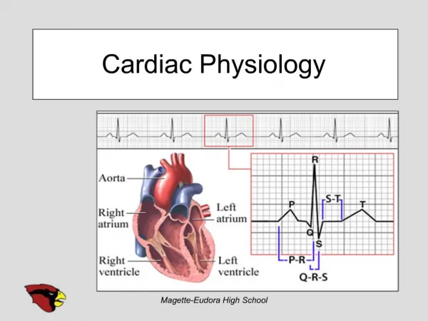
Atrioventricular (AV) Valves • guard the passageway between the atria and the ventricles • Tricuspid valve between right atrium and right ventricle • Bicuspid (mitral) valve between left atrium and left ventricle
Semilunar valves • Between ventricles and arteries • Pulmonary valve between right ventricle and pulmonary artery • Aortic valve between left ventricle and aorta
All myocardial cells • Gap junctions at intercalated discs, waves of depolarization spread from one cell to another
Autorhythmic myocardial cells • (pacemakers) are small myocardial cells with few contractile fibers • Spontaneously generate action potentials • Enables the heart to contract without any outside signal • The heart is myogenic: signal for contraction originates from heart muscle itself
Most myocardial cells • Remaining myocardial cells are striated • Have sarcomeres • Much smaller than skeletal muscle fibers • Connected by gap junctions at intercalated discs • Lots of mitochondria • Lots of blood flow to myocardial cells
More facts about myocardial cells • Large branching t-tubules • Sparse sarcoplasmic reticulum • Source of Ca++ is largely extracellular
Excitation-contraction coupling • Depolarization cell membrane voltage gated Ca++ channels open • Ca++ enters cell • calcium-induced calcium release: Ca++ released from SR • Ca++ binds to troponin contraction
Myocardial cell relaxation • Ca++ dissociates from troponin • Ca++ returns to SR by Ca++ ATPase • Ca++ also transported from cell by Na+-Ca++ indirect active transport protein: Ca++ is exchanged for Na+, which moves in along its electrochemical gradientNa+ removed by active transport
Regulation of cardiac muscle contraction • Graded contractions • Effect of cardiac muscle stretching • Channel activity during action potentials • In myocardial contractile cells • In autorhythmic pacemakers
Graded contraction • The amount of force varies with the number of cross-bridges formed • Low Ca++ few cross-bridges • High Ca++ more cross-bridges
The effect of epinephrine and norepinephrine of contraction • NE and E bind to beta 1 receptors on contractile myocardial cells • The beta 1 receptor is coupled to a G protein • Cyclic AMP is formed
The effect of epinephrine and norepinephrine of contraction • cyclic AMP is formed • 1. Voltage gated Ca++ channels are phosphorylated stay open longer more intracellular Ca++ stronger contractions • 2. A regulatory protein, phospholamban, is phosphorylated increased activity on SR Ca++ ATPase contractions shorten duration
Effect of phospholamban on Ca++ release • NE and E activity • increase phospholamban activity • increase Ca++ ATPase activity on SR • more Ca++ is sequestered into the SR • more Ca++ is available for Ca++ release during stimulation • stronger force of contraction
Effect of NE and E on contraction • Stronger, more frequent contractions
When myocardial cells elongate • The amount of Ca++ entering the myocardial cells may increase the force of contraction increases
Myocardial contractile cell action potentials • Resting potential is stable -90 mV • Wave of depolarization through gap junctions • Voltage gated Na+ channels open • Voltage gated K+ channels open • Slow voltage gated Ca++ channels open and K+ channels close • Ca++ channels close and K+ channels open
Long action potential • Myocardial cell refractory period and contraction end simultaneously
Action potentials in myocardial autorhythmic cells • The channels: • If channels allow passage of Na+ and K+ • Ca++ channels
Action potentials in myocardial autorhythmic cells • Unstable resting membrane potential • Pacemaker potential • At a membrane potential of -60 mV Na+ enters through the If channels • mb depolarizes • Ca++ channels open • Ca++ channels close • K+ leaves
Modulation of autorhythmic cells • NE (sympathetic) and E (adrenal hormone) • Autorhythmic cells have beta1 receptors • Cyclic AMP levels increase • Properties of If and Ca++ channels altered • More rapid Na+ and Ca++ entry • Rapid action potential • Rapid contractions
Modulation of autorhythmic cells • Parasympathetic, acetyl choline • Muscarinic receptors • K+ channels open mb hyperpolarizes cell less excitable • Ca++ channel less likely to open slower depolarization cell is less excitable