Chapter 22
Dive into the intricate world of the lymphatic and immune systems, understanding lymph flow, lymphoid tissues, and the key cells involved in immunity. Discover the functions of lymph nodes and organs like the spleen and thymus.

Chapter 22
E N D
Presentation Transcript
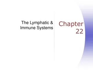
The Lymphatic & Immune Systems Chapter 22
Consists of two semi-independent parts • A meandering network of lymphatic vessels • Lymphoid tissues and organs scattered throughout the body • Returns interstitial fluid and leaked plasma proteins back to the blood • Lymph – interstitial fluid once it has entered lymphatic vessels Lymphatic System: Overview
A one-way system in which lymph flows toward the heart • Lymph vessels include: • Microscopic, permeable, blind-ended capillaries • Lymphatic collecting vessels • Trunks and ducts Lymphatic Vessels
Similar to blood capillaries, with modifications • Remarkably permeable • Loosely joined endothelial minivalves • Withstand interstitial pressure and remain open • The minivalves function as one-way gates that: • Allow interstitial fluid to enter lymph capillaries • Do not allow lymph to escape from the capillaries Lymphatic Capillaries
Lymphatic Vessels and Valves Figure 22–3
The lymphatic system lacks an organ that acts as a pump • Vessels are low-pressure conduits • Uses the same methods as veins to propel lymph • Pulsations of nearby arteries • Contractions of smooth muscle in the walls of the lymphatics Lymph Transport
Lymphocytes are the main cells involved in the immune response • The two main varieties are T cells and B cells Lymphoid Cells
T cells and B cells protect the body against antigens • Antigen – anything the body perceives as foreign • Bacteria and their toxins; viruses • Mismatched RBCs or cancer cells Lymphocytes
T cells • Manage the immune response • Attack and destroy foreign cells • B cells • Produce plasma cells, which secrete antibodies • Antibodies immobilize antigens Lymphocytes
Macrophages – phagocytize foreign substances and help activate T cells • Dendritic cells – spiny-looking cells with functions similar to macrophages • Reticular cells – fibroblastlike cells that produce a stroma, or network, that supports other cell types in lymphoid organs Other Lymphoid Cells
Lymphoid Nodules Figure 22–6
Lymph nodes are the principal lymphoid organs of the body • Nodes are imbedded in connective tissue and clustered along lymphatic vessels • Aggregations of these nodes occur near the body surface in inguinal, axillary, and cervical regions of the body Lymph Nodes
Their two basic functions are: • Filtration – macrophages destroy microorganisms and debris • Immune system activation – monitor for antigens and mount an attack against them Lymph Nodes
Range from 1–25 mm diameter Lymph Nodes Figure 22–7
The spleen, thymus gland, and tonsils • Peyer’s patches and bits of lymphatic tissue scattered in connective tissue • All are composed of reticular connective tissue and all help protect the body • Only lymph nodes filter lymph Other Lymphoid Organs
Innate (nonspecific) system responds quickly and consists of: • First line of defense – intact skin and mucosae prevent entry of microorganisms • Second line of defense – antimicrobial proteins, phagocytes, and other cells • Inhibit spread of invaders throughout the body • Inflammation is its hallmark and most important mechanism Immunity: Two Intrinsic Defense Systems
Adaptive (specific) defense system • Third line of defense – mounts attack against particular foreign substances • Takes longer to react than the innate system • Works in conjunction with the innate system Immunity: Two Intrinsic Defense Systems
The 7 Nonspecific Defenses Figure 22–10
Skin, mucous membranes, and their secretions make up the first line of defense • Keratin in the skin: • Presents a formidable physical barrier to most microorganisms • Is resistant to weak acids and bases, bacterial enzymes, and toxins • Mucosae provide similar mechanical barriers Surface Barriers
Mucus-coated hairs in the nose trap inhaled particles • Mucosa of the upper respiratory tract is ciliated • Cilia sweep dust- and bacteria-laden mucus away from lower respiratory passages Respiratory Tract Mucosae
The body uses nonspecific cellular and chemical devices to protect itself • Phagocytes and natural killer (NK) cells • Antimicrobial proteins in blood and tissue fluid • Inflammatory response enlists macrophages, mast cells, WBCs, and chemicals • Harmful substances are identified by surface carbohydrates unique to infectious organisms Internal Defenses: Cells and Chemicals
Natural Killer Cell Function Figure 22–11
Macrophages are the chief phagocytic cells • Free macrophages wander throughout a region in search of cellular debris • Kupffer cells (liver) and microglia (brain) are fixed macrophages • Neutrophils become phagocytic when encountering infectious material • Eosinophils are weakly phagocytic against parasitic worms • Mast cells bind and ingest a wide range of bacteria Phagocytes
Inflammation is caused by: • Pathogens • Mechanical irritation or damage • Chemical irritants • Extreme temperatures • Marked by four “cardinal signs” • Redness • Pain • Swelling • Heat Inflammation
Vasodilation & increased capillary permeability • Release of: • Histamine – attract leukocytes (chemotaxis), cause further vasodilation and increased permeability • Kinnins – Similar to histamine • Prostaglandins – synergistic with the above. Trigger pain. • Leukotrienes – allow adherence of phagocytes to pathogens • Complement – more histamine, and a bunch of other stuff (about which more later) Overview of inflammatory response
Caused by resetting of hypothalamic thermostat • Bacterial toxins • Triggers release of cytokines & interleukin-1 (endogenous pyrogens) • Increases interferon effects • Increases metabolic rate to enhance tissue repair and increase immune response • Makes environment hostile to microbes Fever
The adaptive immune system is a functional system that: • Recognizes specific foreign substances • Acts to immobilize, neutralize, or destroy foreign substances • Amplifies inflammatory response and activates complement Adaptive (Specific) Defenses
The Immune Response Figure 22–15 (Navigator)
Substances that can mobilize the immune system and provoke an immune response • The ultimate targets of all immune responses are mostly large, complex molecules not normally found in the body (nonself) Antigens
Immature lymphocytes released from bone marrow are essentially identical • Whether a lymphocyte matures into a B cell or a T cell depends on where in the body it becomes immunocompetent • B cells mature in the bone marrow • T cells mature in the thymus Lymphocytes
The MHC is a group of genes that code for proteins that act as an ID badge for your cells • The “self antigens” are glycoproteins that are also called human leukocyte antigens (HLA) • There are two classes, MHC-I & MHC-II • MHC-I is on all cells (except erythrocytes) • MHC-II is found on antigen-presenting cells (APCs) Major Histocompatability Complex and recognition of “self”
Antigen Presentation Figure 22–16b
Helper T Cells (CD4) • Secrete interleukin-2 (IL-2) • Helps activate cytotoxic T Cells, B Cells and/or NK cells • Involved in both cell and antibody mediated responses Types of T Cells
Cytotoxic T Cells (CD8) a.k.a. “killer T Cells” • Attack virally or bacterial infected cells and cancerous cells directly • Memory T Cells provide for a prolonged protection against specific antigens (immunity in the true sense) Types of T Lymphocytes
Cytotoxic T Cell activity:T cell receptor binding, perforin and lymphotoxin release