Download
1 / 12
120 likes | 182 Views
Learn about Chromoblastomycosis caused by dematiaceous fungi and Mycetoma (Maduromycosis) by filamentous fungi. Understand the etiologic agents, clinical manifestations, diagnostic methods, and treatment options.

E N D
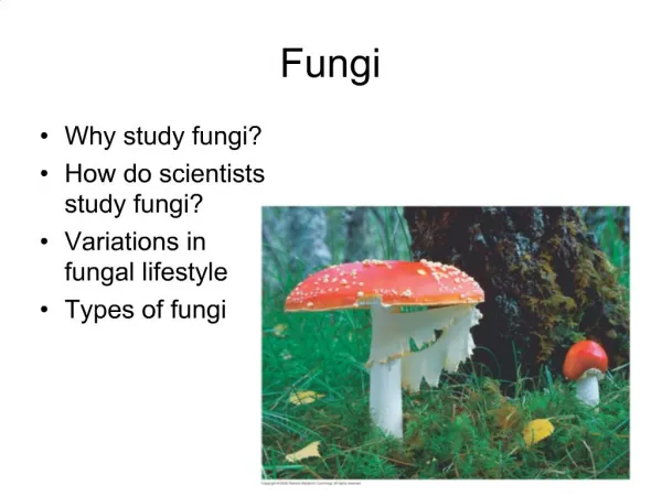
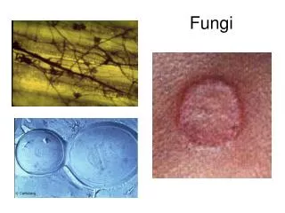
FILAMENTOUS FUNGI A. CHROMOBLASTOMYCOSIS A chronic, localized infection of subcutaneous tissues caused by several species of dematiaceous fungi. The 3 most common agents are: Fonsecaea pedrosoi Cladosporium carrionii Phialophora verrucosa These fungi, recognized by a variety of names, are saprobes located in soil and decaying vegetation. The route of entry is usually by trauma. The lesions are sub-cutaneous and the surface can be flat or verrucous. The lesions take several years to develop.
These organisms are called dematiaceous fungi, because they have a black color in the mycelium cell wall (in culture and in tissue). In tissue these fungi form scleroticbodies which are the reproductive forms dividing by fission. These organisms induce a granulomatous reaction. The etiologic agents of chromoblastomycosis are septate, mold-like, branching, darkly pigmented which produce asexual fruitscalled conidia. We identify these fungi in culture by the shape and formation of the conidia.
The fungi have a world-wide distribution especially in warmer climates like the tropics or the southern U.S. The melanin in the pigment may be a virulence factor. These organisms are distributed world-wide. There is no really successful therapy. Excision and local heat have been used with some success. Flucytosine (5-FC), thiabendazole and itraconazole have also been used to treat (or control) this disease. There are no serological tests to aid in the diagnosis.
B. MYCETOMA (Maduromycosis) Mycetomas (fungous tumors) are also chronic, subcutaneous infections. These are called eumycotic mycetoma (tumors caused by the TRUE fungi as opposed to those caused by actinomycetes). These tumors frequently invade contiguous tissue, particularly the bone. A diagnosis of the etiologic agent is essential for patient management because the prognosis and therapy differs. Mycetoma characteristics: tumefaction - swelling granules - a variety of colors (white, brown, yellow, black) draining sinus tracts
The 3 most common etiologic agents are: Madurella mycetomatis *Exophiala species *Pseudallescheria boydii *The most common in the US. These organisms are associated with the soil, thus you see many infections in the feet and legs. Clinical specimens for diagnosis: pus - with granules tissue – for histological examination
Identification The color, size and texture of the granules are an aid in the diagnosis of mycetomas. The agents of mycetoma are all filamentous fungi which require 7-10 days for visible growth on the culture media and then another several days for specific identification. These fungi are identified by the colonial morphology, conidia formation and biochemical reactions. The species of fungi cannot be distinguished in histopathological tissue sections. Treatment Is very difficult, but ketoconazole and itraconazole have been used with some success.