Download

1 / 23
240 likes | 428 Views
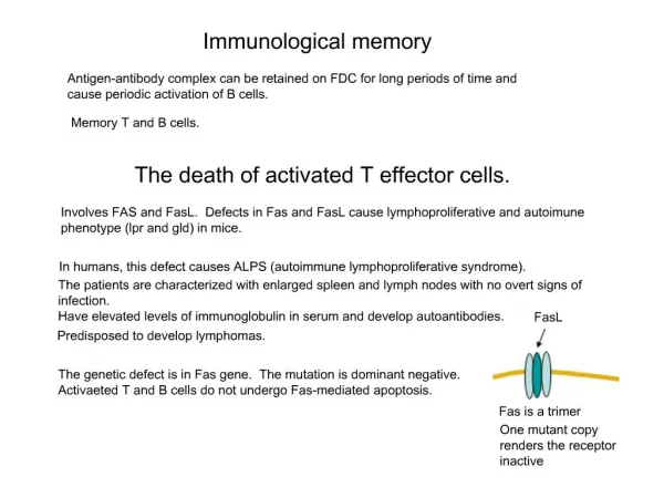
Memory T cells. What is immunological memory?. The capacity of immune cells to “remember” past infections. Which immune cells have the capacity to remember? . Specialized cells known as memory B and T lymphocytes. Why it is important to understand the generation of memory lymphocytes?.

E N D
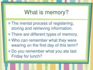
Memory T cells What is immunological memory? The capacity of immune cells to “remember” past infections Which immune cells have the capacity to remember? Specialized cells known as memory B and T lymphocytes Why it is important to understand the generation of memory lymphocytes? Immunological memory is the basis of vaccination. The ultimate goal of a vaccine is to develop long-lived protection, whereby the first encounter with a pathogen is “remembered”, which leads to enhanced memory responses that either completely prevent infection or greatly reduce the severity of disease.
Memory T cells • In this lecture, we will focus on memory T cells , and in particular on CD8+ T cell memory. • We will examine: • Characteristics of memory T cells • Models proposed for the generation of memory T cells • Subsets of memory T cells
Characteristics of Memory T cells What is the essential characteristic of memory T cells? Memory T cells respond more rapidly and more aggressively than naïve T cells. What is the physiological basis for the faster response of memory T cells? • Increased number. Memory T cells are present in higher numbers than naïve T cells. The frequency of a • given antigen-specific T cells in an immune animal can be 1000X higher than in a naïve animal. • Gene-expression profile which is reprogrammed by changes in chromatine structure. For example, mRNA • for IFN-g and cytotoxic molecules such as perforin and granzyme B are not found in naïve T cells • whereas these transcripts are elevated in memory CD8+ T cells. Therefore, memory CD8+ T cells • have the capacity to produce larger quantities of these effector proteins more rapidly than naïve T cells. • Anatomical location. Different pattern of expression of cell surface proteins involved in cell adhesion and • chemotaxis that allow them to gain access to non-lymphoid tissues, the sites of microbial entry.. • Longevity. Memory T cells are maintained for a long time due to antigen-independent homeostatic • proliferation.They are able to maintain their number by continual low-level proliferation (like a stem cells) • in the absence of Ag. The cytokines IL-2, IL-7 and IL-15 are involved in the homeostatic proliferation of • memory T cells. The longevity of memory T cells explain how they can confer long-term protective • immunity.
Stages of a T cell response The kinetics of a first T cell response to a pathogen can be divided into three distinct phases. The first is expansion, in which antigen-specific lymphocytes are activated to divide. The number of lymphocytes rapidly becomes enormous, and the lymphoid organs (such as the spleen and lymph nodes) enlarge to accommodate them. The activated T cells then begin to take on 'effector' functions; they secrete cytokines and kill infected target cells. The second phase is contraction and occurs soon after the pathogen is cleared. Over 95% of the antigen-specific T cells then die. Finally comes the memory phase, in which those T cells that have been spared by the contraction phase survive for long periods, forming a stable pool of 'memory' cells. Figure 1 | Antiviral CD8+ and CD4+ T-cell responses. The three phases of the T-cell immune response (expansion, contraction and memory) are indicated. Antigen-specific T cells clonally expand during the first phase in the presence of antigen. Soon after the virus is cleared, the contraction phase ensues and the number of antigen-specific T cells decreases due to apoptosis. After the contraction phase, the number of virus-specific T cells stabilizes and can be maintained for great lengths of time (the memory phase). Note that, typically, the magnitude of the CD4+ T-cell response is lower than that of the CD8+ T-cell response, and the contraction phase can be less pronounced than that of CD8+ T cells. The number of memory CD4+ T cells might decline slowly over time.
Models of memory T cell differentiation The divergent model proposes that a naïve T cell can give rise to daughter cells that develop into either effector or memory T cells. Accordingly, naïve T cells can bypass an effector-cell stage and develop directly into memory T cells.
Earlier discovery: a green fluorescent protein reporter gene (T-GFP)is expressed in naive and short-term activated T cells but is silentin terminally differentiated effector cells. In T-GFP transgenic mice,GFP expression can be conveniently used to monitor T cell differentiation. Manjunath et al. have generated naive T cells from T-GFPmice carrying a class I–restricted T cell receptor (TCR)that recognizes a specific viral peptide. They stimulatedthese cells in vitro with antigen for 2 days and expanded themin the presence of different cytokines. When cultured in highdoses of IL-2 (CD8IL-2), the cells become large blasts, expresshigh levels of activation markers, lose expression of GFP and acquire the capacity to produceIFNg- and to kill target cells. The same cells culturedin the presence of low doses of IL-2 or with IL-15 (CD8IL-15),become small, retain GFP, and fail to acquirecytotoxic function, although they acquire IFNg–producingcapacity. Importantly, after adoptive transfer, CD8IL-15 cellssurvive for several weeks and, upon antigen rechallenge, mounta secondary response that is comparable to that mediated byendogenously generated memory cells. Effector differentiation is not prerequisite for generation of memory cytotoxic T lymphocytes N. Manjunath et al. J. Clin. Invest. 108, 871-878 (2001)
Models of memory T cell differentiation According to the linear differentiation model, memory T cells are the progeny of effector T cells. There are many in vitro and in vivo studies that have established that long-term memory results when naïve T cells are induced to undergo several rounds of cell division in response to antigen in vitro and are then adoptively transferred in vivo in the absence of antigen. For example, Opferman et al. demonstrated that only effector T cells that have divided more than five times in vitro can generate memory T cells. Linear differentiation of cytotoxic effectors into memory T lymphocytes Joseph T. Opferman, Bertram T. Ober, Philip G. Ashton-Rickardt Science283, 1745- 1748 (1999)
Models of memory T cell differentiation The decreasing potential hypothesis. The previous model does not definitely resolve the issue of whether memory cells arise from fully differentiated effectors. It is notable that memory fails to occur when T cells undergo `exhaustive' proliferation to high doses of viruses; in this situation, effector T cells are generated in enormous numbers but then die en masse, presumably because the cells are all driven to a terminal stage of differentiation where they can not escape from `activation-induced cell death' (AICD). This phenomenon is known as clonal exhaustion. Thus, cumulative encounter with Ag increases susceptibility of effector T cells to apoptosis and reduced formation of memory T cells.
Thus, increasing cell stimulation and division are associated with progress to terminal differentiation and a reduction in the memory potential. In other words, cells that divide many times are more likely to die than to survive as memory cells. Accordingly, the decreasing potential hypothesis proposes that memory cells may normally arise from a subset of cells that express a full range of effector functions but, perhaps because of lack of prolonged contact with antigen, do not initiate AICD. Hence memory cells might originate from a population of effector cells that only arrive during the later stages of the immune response, when the Ag is removed or greatly decreased in concentration.
Thus, the mechanisms involved in the generation of memory T cells remain poorly understood despite that the practice of “variolation” (inoculation of virus taken from pustules of smallpox victims) was used for protection against smallpox well before 1796. Part of this deficiency may arise from the fact that memory T cells are heterogeneous.
Subsets of Memory T cells Based on the expression of CCR7 two subsets of memory T cells have been recently identified. Figure 1 CCR7 and CD62L are co-expressed on a subset of peripheral blood memory CD4+ and CD8+ T cells. CD4+ (a, b) and CD8+ (c, d) lymphocytes were stained with monoclonal antibodies to CD45RA and CCR7, which identified three and four subsets, respectively. These subsets were sorted and analysed for the expression of CD62L, and the percentage of bright cells is indicated (b, d). Upon serial analysis, the proportion of cells in the different compartments was rather stable in the same individual, but more variable among individuals, the variability being more pronounced in the CD8 than in the CD4 compartment. Comparable results were obtained using two anti-CCR7 antibodies (clones 3D12 and 10H5). Two subsets of memory T lymphocytes with distinct homing potentials and effector functions FEDERICA SALLUSTO*, DANIELLE LENIG*, REINHOLD FÖRSTER†, MARTIN LIPP† & ANTONIO LANZAVECCHIA* Nature401, 708 - 712 (1999)
Figure 2 CCR7+ and CCR7- memory T cells display different effector functions.c, d, The four subsets of CD8+ T cells were sorted according to the expression of CCR7 and CD45RA as in Fig. 1 and tested for their capacity to produce IL-2 or IFN- (c) or were immediately stained with anti-perforin antibody (green) and counterstained with propidium iodide (red) (d). In the CD8+ CD45RA+ compartment, CCR7 expression allows us to discriminate naive cells (1) from effector cells (4) (ref. 26). Comparable results were obtained in 12 healthy donors. Memory CCR7+ Naive Memory CCR7- Effectors Two subsets of memory T lymphocytes with distinct homing potentials and effector functions FEDERICA SALLUSTO*, DANIELLE LENIG*, REINHOLD FÖRSTER†, MARTIN LIPP† & ANTONIO LANZAVECCHIA* Nature401, 708 - 712 (1999)
Two subsets of memory T cells: CCR7+ CD62Lhigh Central memory T cells (CM) CCR7- CD62Llow Effector memory T cells (EM) With functional differences: TCM IL-2, little IFN-g, no perforin TEM little IL-2 but high IFN-g and perforin And different homing potential: CD62L interacts with PNAd on HEV, which mediates attachment and rolling. CCR7 binds to chemokines CCL19 and CCL21 that are presented on the luminal surface of endothelial cells in lymph nodes which causes firm arrest and the initiation of extravasation. Studies have shown that CCR7+ CD62Lhigh T cells migrate efficiently to peripheral lymph nodes, whereas T cells lacking these two molecules do not. Rather, CCR7- CD62Llow T cells can be found in other sites, such as the liver and lungs.
Masopust et al. tracked the migration of CD8+ memory T cells with tetramers composed of major histocompatibility complex (MHC) molecules bound to an antigenic peptide. Their tetramer was composed of mouse MHC class I molecules and a peptide derived from vesicular stomatitis virus (VSV). This tetramer identified VSV-specific CD8+ T cells in mice that had been infected with this virus. The dynamics and distribution of VSV-specific CD8+ T cells were revealed by analyzing which T cells bound to the tetramer. Remarkably, 9 days after infection, nonlymphoid tissues including kidney, liver, and peritoneum contained extremely high numbers of VSV-specific CD8+ T cells (which constituted up to 40% of the total CD8+ population). Even after 296 days, some tissues still retained VSV-specific T memory cells that constituted as much as 4% of the total CD8+ population. Despite the high proportion of VSV-specific CD8+ cells in nonlymphoid tissues, this T cell subpopulation had almost totally disappeared from lymphoid tissues. Thus, the clonally expanded effector and memory T cells had become redistributed to the body's nonlymphoid tissues, the very places where protection against pathogens is needed the most. Preferential localization of effector memory cells in nonlymphoid tissue David Masopust, Vaiva Vezys, Amanda L. Marzo, Leo Lefrancois Science291, 2413- 2417 (2001)
Figure 1. Infection with VSV leads to the appearance of virus-specific CD8 T cells in lymphoid and nonlymphoid tissues. C57Bl/6J mice were infected intravenously with 106 plaque-forming units (PFU) of VSV-Indiana, and 8 days (A) or 81 days (B) later, mice were perfused and lymphocytes were isolated from the indicated tissues. The percentage of antigen-specific CD8 T cells was assessed by staining with N52-59/Kb tetramer and antibodies to CD8 and CD11a, followed by fluorescence flow cytometry. Plots shown are gated on CD8+ lymphocytes; values are mean percentages of tetramer+ cells within the CD8+ T cell population derived from at least four mice. Control staining with a Kb tetramer containing SIINFEKL was negligible. PLN, peripheral lymph nodes; MLN, mesenteric lymph nodes; PBL, peripheral blood lymphocytes; LP, small intestine lamina propria; IEL, small intestine intraepithelial lymphocytes; BM, bone marrow; Perit, peritoneal cavity lymphocytes.
They also demonstated that the T cells redistributing to nonlymphoid tissues have an activation profile characteristic of Sallusto's effector-memory T cells. In contrast, lymphoid memory T cells, that did not have Immediate effector ability resembles Sallusto's central-memory T cells. Fig. 3. Virus-specific CD8 memory T cells in peripheral but not lymphoid tissues are constitutively cytolytic. (B) Twenty days after VSV infection, lymphocytes were incubated for 4 to 5 hours with 51Cr-labeled untreated EL4 target cells (27) or target cells pulsed with N52-59 peptide. E:T ratio was 200:1 for all tissues. E:T values shown in plots are corrected for the number of tetramer+ cells in each population. (C) As described in (B), except lymphocytes were isolated from mice primed with 106 PFU of VSV-New Jersey, rested >7 months, then infected with VSV-Indiana and rested an additional 224 days. E:T ratio was 300:1 for all tissues. E:T values shown in plots arecorrected for the number of tetramer+ cells Preferential localization of effector memory cells in nonlymphoid tissue David Masopust, Vaiva Vezys, Amanda L. Marzo, Leo Lefrancois Science291, 2413- 2417 (2001)
Reinhardt et al.reached a similar conclusion although they tracked the redistribution of CD4+ T cells using a totally different approach. They followed migrating antigen-specific CD4+ T cells by transferring naïve T cells with a defined antigenic specificity (derived from a T cell receptor transgenic mouse) into recipient mice and using Thy-1 as a marker for the transferred cells. The authors painstakingly determined the presence of antigen-reactive CD4+ T cells in all tissues of the recipient animals. They did this by immunohistochemical analysis of whole-body sections using an antibody against Thy-1.1 that only bound to donor-derived T cells. With this protocol, they first tracked the migration of transferred naïve T cells, and then monitored changes in their migration after injection of the specific antigen. As expected, naïve T cells initially became localized only within lymphoid tissues. However, after injection of antigen and a primary immune response, there was a striking redistribution of antigen-reactive T cells to nonlymphoid tissues including the liver, lungs, and intestinal lamina propria. This study also demonstrated that memory T cells migrating to nonlymphoid tissues can rapidly become effector cells. Visualizing the generation of memory CD4 T cells in the whole body R. LEE REINHARDT*, ALEXANDER KHORUTS†, REBECCA MERICA*, TRACI ZELL* & MARC K. JENKINS* Nature410, 101 - 105 (2001)
Although, these studies did not address the phenotype of tissue-derived memory T cells with respect to CD62L and CCR7, they have confirmed the presence of antigen-specific memory T cells in non-lymphoid compartments long after priming, which supports the notion of an effector memory subset of T cells. What is the wider implication of these studies? First, a clear distinction can be made between the migration pathways of naïve T cells and those of tissue memory T cells. Second, rapid memory responses occur because antigen-reactive cells are greatly expanded in number and have redistributed to numerous tissues to provide "frontline" immune protection. But there is more to immunological memory than simply an increase in the number of antigen-reactive cells. These studies demonstrated that memory T cells migrating to nonlymphoid tissues can rapidly become effector cells. A model was proposed in which the tissue-homing effector memory T cells, which are capable of immediate effector functions, could rapidly control invading pathogens. TEM= first line of defense in tissues The lymph-node-homing central memory T cells would be available in secondary lymphoid organs ready to stimulate dendritic cells, provide B-cell help and/or generate a second wave of T-cell effectors. TCM= reserve of defenses
The major question resulting from these findings is how these two subsets of memory T cells are generated. Three models of differentiation have been proposed: Two subsets of memory T lymphocytes with distinct homing potentials and effector functions FEDERICA SALLUSTO*, DANIELLE LENIG*, REINHOLD FÖRSTER†, MARTIN LIPP† & ANTONIO LANZAVECCHIA* Nature401, 708 - 712 (1999) In vitro stimulation of naive T cells resulted in the generation of both TCM and TEM cells, whereas stimulation of TCM cells resulted in their efficient differentiation to TEM cells. These data were consistent with a linear differentiation model in which naive T cells differentiate first to TCM and then to TEM cells, which were considered end-stage cells. Naive effectorsTCM TEM
+ + Migratory properties of naive, effector, and memory CD8(+) T cells Weninger W, Crowley MA, Manjunath N, von Andrian UH. J. Exp. Med. 194, 953-966 (2001) According to Manjunath et al., the duration of antigenic stimulation and the type and amount of cytokines present during priming lead either to fully differentiated effector cells that home to peripheral tissues (blue) or to cells that are devoid of effector function and home to lymph nodes (green). In the system used by Manjunath et al., these two cell types can be identified according to the differential expression of the T-GFP marker transgene and the lymph node–homing receptor CCR7. Both cell types are maintained in the memory pool (dotted arrows) and, upon secondary challenge, mediate immediate protection in nonlymphoid tissues or secondary responses in lymph nodes. The repertoires of circulating human CD8+ central and effector memory T cell subsets are largely distinct. Baron V., Bouneaud C., Cumano A., LimA., Arstila T.P., KourilskyP., FerradiniL.,and Pannetier C. Immunity 18, 193-204 (2003) Baron et al. analyzed the composition and dynamics of the CD8+ T cell repertoire of these subsets within the peripheral blood of four healthy individuals. Both subsets had largely distinct and autonomous TCRV repertoires. Their composition remained stable over a 9 month period, during which no cell passage between these subsets was detected despite important size variation of several clones. In one donor, four out of six TCRV clonotypes specific for the influenza A virus were detected in the central subset only, while the two others were shared. Altogether, these observations suggest that most effector memory T cells may not have derived from the central memory subset.
Memory B cells • Generated in germinal centers • therefore we only have humoral memory to T-dependent antigens • Small, recirculating cells • Typically isotype switched (e.g. IgG+ or IgA+) • Typically have higher affinity for the inducing Ag • Longer lived than naïve B cells • Persistence of memory B cells after an immune response ensures that we have increased numbers of B cells specific for the antigen and ready to respond on re-encounter