Download
1 / 62
630 likes | 829 Views
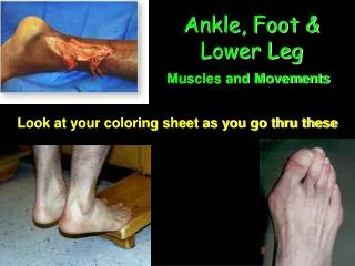
Ankle, Foot & Lower Leg. Look at your coloring sheet as you go thru these. Muscles and Movements. Achilles Tendon. Achilles Tendon. Origin Stems from the distal ends of both the Soleus and Gastrocnemius Muscles Insertion Posterior Surface of the Calcaneus Palpation

E N D
Ankle, Foot & Lower Leg Look at your coloring sheet as you go thru these Muscles and Movements
Achilles Tendon • Origin • Stems from the distal ends of both the Soleus and Gastrocnemius Muscles • Insertion • Posterior Surface of the Calcaneus • Palpation • Very easy to palpate the distal end of the calf to the Calcaneus
Extensor Digitorum Longus • Origin • Lateral Condyle of the tibia, Fibular Head, upper two thirds of anterior fibula • Insertion • Tops of the Distal and Middle 2 - 5 Phalanges • Palpation • 2nd Muscle upper lateral side of tibia / RROM Toe ext dorsal foot
Extensor Digitorum Brevis • Origin • Anterior/Lateral Surface of the Calcaneus • Insertion • Tops of the Distal and Middle 2 - 5 Phalanges • Palpation • Can not
Extensor Hallucis Longus • Origin • Middle 2/3 of medial anterior fibula • Insertion • Top of the distal 1st Phalange • Palpation • RROM Great Toe Ext
Extensor Hallucis Brevis • Origin • Anterior Lateral part of the upper Calcaneus • Insertion • Dorsal surface on the base of the 1st Proximal Phalange • Palpation • Can Not
Flexor Digitorum Longus • Origin • lower two thirds of the posterior tibia • Insertion • Base of the distal phalanx of toes 2-5 • Palpation • Can not palpate
Flexor Digitorum Brevis • Origin • Medial Calcaneal Tuberosity • Insertion • Divides into two heads at the base of the Proximal Phalange • Allowing the FDL to slip through • Attaching to each side of the middle phalange 2-5 • Palpation • Plantar side of the foot with RROM Toe Flexion
Flexor Hallucis Longus • Origin • Lower two thirds of the posterior fibula • Insertion • Base of the distal posterior big toe • Palpation • Down the medial malleolus RROM Big toe flexion
Flexor Hallucis Brevis • Origin • 2 Heads • Plantar surface of the Cuboid & 3rd Cuneiform • Plantar surface of the 2nd and 1st Cuneiforms • Insertion • 2 Heads • One on each side of the Base of the Proximal 1st Phalange • Encompasses a Sesmoid • Palpation • Unable – Irritation with the Sesmoids however
Gastrocnemius • Origin • Posterior surfaces of the two femor condyles • Insertion • posterior surface of calcaneus • Palpation • Easiest muscle to palpate Upper posterior lower leg
Peroneus Longus • Origin • Head and upper thirds of the outer surface of the fibula • Insertion • undersurfaces of the 2nd cuneiform and first metatarsal • Palpation • third muscle on the lateral side of the tibia, upper lateral tibia
Peroneus Brevis • Origin • Lower two thirds of the lateral surface of the fibula • Insertion • Styloid Process • Palpation • Palpate at the Styloid Process
Peroneus Tertius • Origin • Lower third of the lateral surface of the fibula • Insertion • Styloid Process • Palpation • Palpate at the Styloid Process
Motions 5th
Plantaris • Origin • Posterior / Medial side of the Lateral Femoral Condyle • Insertion • Medial / Posterior Surface of the Calcaneus • Palpation • Partially at Insertion Point
Soleus • Origin • Upper two thirds of the posterior surfaces of the tib/fib • Insertion • posterior surface of calcaneus • Palpation • under gastroc on the lateral side of the lower leg
Tibialis Anterior • Origin • Upper two thirds of the lateral surface of the tibia • Insertion • Medial surface of the 1st cuneiform and first met • Palpation • First muscle on the lateral side of the tibia