Download
1 / 13
130 likes | 164 Views
The Michael Hooker Microscopy Facility offers maintenance, user training, workshops, and software updates for various microscopy equipment. They provide support for laser scanning confocals, high-resolution cameras, and image processing software.

E N D
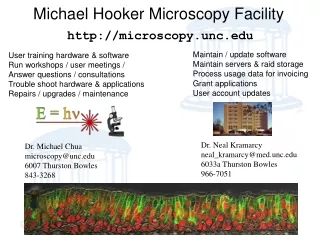
http://microscopy.unc.edu Michael Hooker Microscopy Facility Maintain / update software Maintain servers & raid storage Process usage data for invoicing Grant applications User account updates User training hardware & software Run workshops / user meetings / Answer questions / consultations Trouble shoot hardware & applications Repairs / upgrades / maintenance Dr. Neal Kramarcy neal_kramarcy@med.unc.edu 6033a Thurston Bowles 966-7051 Dr. Michael Chua microscopy@unc.edu 6007 Thurston Bowles 843-3268
Laser Scanning Confocals Leica - SP2 aobs UV 351, 364 nm blue 458, 488 nm green514, 561 nm red 633 nm Reflection Transmitted DIC X-Z scanning l scanning FRAP, FRET Zeiss - 510 meta blue 458, 490 green 514, 543 red 633 nm Reflection Transmitted DIC Spectral unmixing FRAP, FRET Fluorophores: DAPI, Hoechst, Alexa 350, etc FITC, GFP, Alexa 488, etc. Rhodamine, Texas Red, Alexa 543, 568 & 594 CY5, DRAQ 5, Alexa 635 (far red, invisible) CFP/YFP, dsRed,etc.,fluorescent proteins many others… Heated chambers, 5% CO2 Tissues, small animals possible with setup UltraView blue 488 nm green 568 nm red 633 nm transmitted Biosensors
Michael Hooker Microscopy Facility Resources: – Digital Light Microscopy • Instrumentation • Microscopes: 6xCompound, 2x Dissecting, 3x Confocals, Laser Micro-Dissection, Atomic Force, • Flat bed scanner (standard reflected and transparency) • Imaging • Processing, Analysis, morphometry • 2D, 3D, time lapse, FRAP, FRET, Fura-2, tracking • Storage, color printing (Xerox dye sub) • File storage (raid arrays, Ethernet accessible) • Instruction • Advice • Training • Help • Seminars/courses/workshops on microscopy • Collaborations • Fee for use – recharge facility • Credit for block support e.g. PPG, SCOR cores • Users from UNC-CH, EPA, Pharmas, NCCU
Wide Field Microscopes Dissecting Scopes Fluorescent & motorized Simple transmitted light Leica DMIRB inverted Transmitted light Nomarski (DIC) Fluorescence B/W camera (OrcaER) Color camera Nikon TE2000 inverted Transmitted light Nomarski (DIC) Phase contrast Fluorescence Time lapse Ratio imaging e.g. Fura-2 [Ca], FRET Multimode B/W cameras Intensified camera Motorized filters changers Nikon upright Transmitted light Hi resolution color camera
Mission: • Research light microscopy facility • Standard and advanced digital light microscopy image acquisition • Image processing and analysis resources • Provide instrumentation and instruction • Fee for use, training, assistance • Users scan and analyze images from samples that they have prepared • Occasional users receive scanning assistance (no training) Michael Hooker Microscopy Facility
Location: 6129 Thurston Bowles Michael Hooker Microscopy Facility
Laser Micro Dissection Leica AS-LMD Section on penfoil slide (polyethylene coated) Outline area on monitor image Cut with pulsed UV laser Collect cut out in Eppendorf tube cap Protein mass spec. / DNA / RNA analysis Ablation of Zebra fish vasculature
Image Processing Work Stations (4x) \\Nomarski – general purpose – full Leica confocal off line \\Snell – 3D Volocity, full Leica & Zeiss confocal off line \\Zernike – Windows 64 bit RAM 8GByte OpenGL 512MByte Video \\Young – video tape capture – scanner with transparency Resources: Image Analysis & Processing Software (examples understated!) General:- C-Imaging 6, Metamorph 7.1, ImageJ 3D Rendering:- Volocity, Slicer (T3D), Voxblast Volocity licence server http://ai.med.unc.edu:15003 2D:- Photoshop, Illustrator, ThumbsPlus 7 Video:- Quicktime Pro 7, VidEdit 1.1, Premiere 1.5, Analysis Co-localization, fluorograms, FRAP, FRET Measurements – area – intensity – tracking - etc. Enhancements – min/max/ave projections – local contrast – etc. Etc., etc., etc…..
Summary: • Resources: • Space 850 sq.ft • Microscopes 10x • Workstations 4x • Software 3x high packages • Web pages & booking calendars http://microscopy.unc.edu • File servers / AD-DC / Terabyte Raid Windows files/directory sharing • Personnel 2x • What we do: • User Training, on demand – New users $140 confocals, $70 widefields, • Consultations (in person, on demand, e-mail, phone) $9.50 / 15 minutes • Collaborations • User problems $9.50 / 15 mins, but mostly not logged • Image analysis and data processing development $9.50 / 15 mins, somwhat logged • Annual Workshops $350 to $700 5day with discount to those who give salary support/ Users group meetings $free • Support Cystic Fibrosis Center, Alcohol Studies Center, Gene Therapy Center, SukWon Jin lab. (R01) • Director salary • CF: Scor 0.25 ended Aug 2009 • CF: Core-D 0.20, • CF-GT: MTCC 0.10 • GT: PPG 0.1 end Oct 2009 • Alc: P60 0.1 • Jin: RO1 - 0.05 • Recharge: q.v. • Resource maintenance/repairs/trouble shooting/system development, including computer IT • Book keeping/grants/l.o.s. • Olympus Center out reach (No details of activities yet) • What we do not do: • Tissue preparation (but advice given) • Scan for users (some exceptions $9.50 /hr + machine cost) • Short comings: • No wet bench space • No sterile hood (bad for biosensor experiments) • Server room (With 3 servers overnight office temperature is 26oC) • Older equipment approaching end of service life • Have to compete with UCRF supported facilities – i.e. PIs do not want to contribute to shared instrumentation grants. Those cores have lower prices. • Need more core grants with UNC groups
\\Marvin file server 4.6 x 1012 bytes Active Directory Domain Controller \\Minsky file server 1.3 x 1012 bytes Domain Controller \\AI file server 9.4 x 1012 bytes Backup server Shared network resources, e.g: \\MINSKY\USERS2public (any MHMF users) \\MINSKY\USERS1 public & AppleTalk \\MINSKY\PI-lab share: lab. group \\MINSKY\yournameshare: private \\MINSKY\Phaser8550-MHMF share: dye sub color printer \\computername\shareddirectory\filename UNC (Universal Name Convention) Incubator 37oC, 5% CO2, 95% humidity Refrigerator Heated Stages Other Resources
Using the resources: • Training/orientation for each microscope • User are issued with computer account on mhmicroscopy domain • Users may request after hours access • Wet sample restrictions • Booking: • On line http://microscopy.unc.edu/booking/ • Walk up if machine is not booked • Assistance with experiments, consultations, collaborations: • Schedule an appointment • Ad Hoc if less than 15 minutes • (Please do not assume we are available without an appointment) Using: The Resources
http://microscopy.unc.edu ftp://152.19.58.173