Download
1 / 24
240 likes | 419 Views
UofR : Neural Basis of Cognition Lecture 2. Methods, Part II Hemispheric Specialization 25 May 2010. Electromagnetic Recording Methods. Electromagnetic Recording Methods Electroencephalography (EEG) Event-Related Potentials (ERPs) Magnetoencephalography (MEG) Optical Recording Methods.

E N D
UofR: Neural Basis of CognitionLecture 2 Methods, Part II Hemispheric Specialization 25 May 2010
Electromagnetic Recording Methods • Electromagnetic Recording Methods • Electroencephalography (EEG) • Event-Related Potentials (ERPs) • Magnetoencephalography (MEG) • Optical Recording Methods
Electroencephalography (EEG) • Method of recording the brain’s electrical activity • Metal electrodes are evenly spaced on the scalp • Each electrode records the electrical potential, the sum of electrical neural activity, where it is placed, and amplifies it (units: Hertz, Hz)
Electroencephalography (EEG) • The electrical potential under every electrode oscillates • Brain activity: • Alpha: 9 – 12 Hz, predominates when relaxed with eyes closed • Alpha suppression: indicator of degree of activation in the brain • Beta: ~15 Hz, predominates while awake and alert • Delta: 1 – 4 Hz, predominates during sleep • Gamma: 25 – 100 Hz, unknown role
Electroencephalography (EEG) • Normally, neurons fire in a synchronized manner (alpha/beta/delta waves) • Epilepsy: neurons fire in bursts at random times
Event-related potentials (ERPs) • Recordings of the brain’s activity that are linked to the occurrence of some event • Gives an idea of “when” processes occur in the brain • As time passes after a stimulus, the active group of neurons changes and the EEG waveforms change accordingly • The waveform can be divided into components, characteristic portions of the wave that have been linked to certain processes
Event-related potentials (ERPs) • ERP components are usually given names: a letter and a subscripted number; the letter is either P (for positive) or N (for negative) • Exogenous components are linked to the physical characteristics of a waveform and occur early in the waveform • Endogenous components appear to be independent of stimulus characteristics and to be driven by internal cognitive states
Event-related potentials (ERPs) • <100ms: sensory processing; an abnormality in early ERPs indicates a disruption in the sensory system • ~100ms: no longer driven solely by sensory information, modulated by attention • N2 (200-300ms): Mismatch negativity • P3 (300-800ms): related to attention and updating of memory (“oddball paradigm”) • N4 (400-600ms): “Running out the door, Patty grabbed her hacker, her baseball glove, her cap, a softball, and a lamp.”
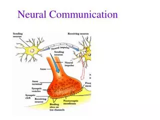
Magnetoencephalography (MEG) • Records induced magnetic potentials instead of electric potentials, allowing location of activity to be pinpointed • Uses superconductors • Used to localize source of epileptic activity, to locate primary sensory cortices to be avoided in surgery, and for more general studies such as of schizophrenia • Example: M100 stimulus generated in atypical location in Heschl’sgyrus, supporting that paranoid schizophrenics have difficulties in filtering early sensory information properly
Techniques for Analyzing Behavior • Test batteries • Estimate of Premorbid Functioning
Computational Neuroscience • Neural Network • Input, hidden, output neurons • Synaptic weights • Use in computer science • Hebbian processes • Perceptron: single-layer neural network
Hemispheric Specialization • First evidence: Broca’s area, 1860s • Right frontal lobe: extends farther forward, is wider • Left occipital lobe: extends farther back, is wider • Sylvian fissure: for most people, extends father in the horizontal direction in the left, takes more of an upward turn on the right • Brodmann’s area 29 is larger on the left • More NE in certain regions of right thalamus, more DA/DARs in the basal ganglia on the left
Hemispheric Specialization: Methods • Divided visual field technique • LVF/RVF project exclusively to the contralateral hemisphere • Unilateral presentation • Bilateral presentation to test perceptual asymmetries • Dichaptic presentation • Analogous but for tactile modality: objects placed in both hands simultaneously and a person is asked to somehow identify/distinguish the items
Hemispheric Specialization: Methods • Dichotic presentation • Analogous but for auditory input • Different auditory information is given to each ear • Competition between inputs leads to suppression of ipsilateral sensory information all\most entirely in favor of contralateral sensory information in each ear
Hemispheric specialization: Findings • Visual: • RVF/left hemisphere advantage for words • LVF/right hemisphere advantage for faces • Tactile: • Right hand/left hemisphere advantage for identifying letters drawn on the palm • Left hand/right hemisphere advantage for identifying complex • Auditory: • Right ear/left hemisphere advantage for responding to words • Left ear/right hemisphere advantage for responding to non-linguistic stimulus • To grossly generalize, the left hemisphere is “verbal” and the right is “nonverbal”
Split-Brain syndrome • Corpus callosum is critical in interhemispheric communication • Split-brain procedure for epilepsy • Objects could verbally identify objects by touch only if they were held in the right hand • Chimeric images • http://www.youtube.com/watch?v=ZMLzP1VCANo
Interhemispheric Communication • Simple tasks are performed faster intrahemispherically • More complex tasks are performed faster interhemispherically
Interhemispheric Communication • Corpus callosum: 250m fiber topographic line of communication between the hemispheres • There are other subcorticalcommissures but they are not as important • For example, callosotomy patients cannot determine whether faces presented in each of their visual fields are the same person • Some information can be transferred, e.g. whether the face was old or young • Callosal transfer time: 5-20ms
Individual Differences in Brain Organization • Handedness • 90% of population is right-handed • Population of right-handed people: • 95% have speech output controlled by left hemisphere • 5% by right • Population of left-handed people: • 70% have speech output controlled by right hemisphere • 15% by right • 15% by both
Individual Differences in Brain Organization • Gender • Difficult to determine gender-specific aspects of brain function • Little to no conclusive evidence of anything apart from brain size