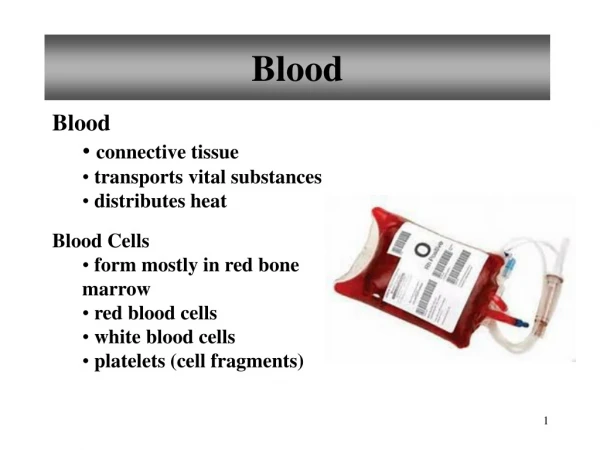
Download
1 / 16
210 likes | 637 Views
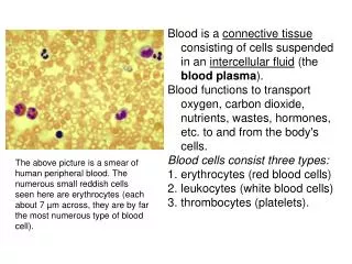
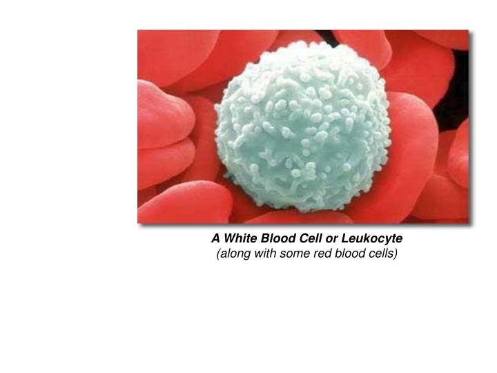
A White Blood Cell or Leukocyte (along with some red blood cells). Clinical Examples of Leukocyte - Induced Injury Acute • Acute respiratory distress syndrome • Acute transplant rejection • Reperfusion injury • Septic shock • Vasculitis. Chronic • Arthritis • Asthma

E N D
A White Blood Cell or Leukocyte(along with some red blood cells)
Clinical Examples of Leukocyte- Induced Injury Acute • Acute respiratory distress syndrome • Acute transplant rejection • Reperfusion injury • Septic shock • Vasculitis Chronic • Arthritis • Asthma • Atherosclerosis • Glomerulonephritis • Chronic lung disease • Chronic rejection
LeukocyteFunctional Defects/Defecits • ►leukocyte functional defects, both genetic and acquired, lead to increased vulnerability to infections: • 1. Inadequate number • 2. Inadequate functioning due to defects in: • – adhesion • – chemotaxis • – phagocytosis / phagolysosome formation • – microbicidal activity
Defects in LeukocyteFunction • Genetic • ●Leukocyte adhesion deficiency 1 • • β chain of CD11/CD18 integrins • ●Leukocyte adhesion deficiency 2 • • Fucosyl transferase required for synthesis of sialylated • oligosaccharide (receptor for selectin) • ●Chronic granulomatous disease • • Decreased oxidative burst • – X-linked • • NADPH oxidase (membrane component) • – Autosomal recessive • • NADPH oxidase (cytoplasmic components) • ● Myeloperoxidase deficiency • • defectiveMPO-H2O2 system • ●Chédiak-Higashi syndrome • • Protein involved in organelle membrane fusion
Defects in LeukocyteFunction • ●Chronic granulomatous disease (CGD) • • CGD is caused by a defect or a deficiency in phagocytic NADPH oxidase, resulting in absence or inadequacy of hydrogen peroxide production. • • This leads to recurrent life-threatening bacterial and fungal infections –most commonly in lungs, skin, and GI. CGD refers to the characteristic granulomas that develop in response to chronic inflammation. • • median survival duration is about 20-25 years. • (There has been increasing success with bone marrow stem cell transplant.) Chronic granulomatous disease with nodular lesions of the face.
●Chronic granulomatous disease (CGD) (cont.) • – X-linked (60-70% of all cases; typically associated with more serious disease) – the most common molecular defect in CGD is a mutation in the CYBB (cytochrome B - b subunit, or gp91) gene. More than 350 mutations in the CYBB gene have been identified. • – Autosomal recessive (20-30% of all cases) • • NADPH oxidase (cytoplasmic components) • ♦In general, carriers of CGD are asymptomatic. However, carriers of X-CGD have a notable incidence of discoid lupus erythematosus, photosensitivity, Raynaud phenomenon, and aphthous ulcers.
Defects in LeukocyteFunction ● Myeloperoxidase deficiency Myeloperoxidase (MPO) catalyzes the conversion of hydrogen peroxide and chloride ions into hypochlorous acid. Hypochlorous acid is 50 times more potent in microbial killing than hydrogen peroxide. ♦ Acquired MPO deficiency -- usually partial and transient (generally resolves once the inciting condition improves). -- conditions which can lead to acquired MPO deficiency = Pb toxicity, Fe deficiency, thrombotic disease, diabetes mellitus, leukemias, some hematologic disorders, some antineoplastic drugs. ♦ Hereditary MPO deficiency -- most patients are compound heterozygotes. ◊ Incidence rate =1 in 1500 population ◊ Morbidity: -- most individuals with partial or total MPO deficiency have no increased frequency of infections, probably because MPO-independent mechanisms in the PMNs can take over. If severe infectious disease occurs, it usually is a fungal infection. These primarily occur in a patient who also has diabetes mellitus.
Defects in LeukocyteFunction Disseminated Candida albicans infection in a patient with hereditary myeloperoxidase deficiency. A 52 year old female presented with a fever of 39°C, painful hyperpigmented skin lesions, malaise, sore mucosae and dysphagia of 1 week's duration. There were palpable painful large inflammatory nodules in the involved skin areas. Laboratory findings were ESR 127/132, Ht 39%, WBC 23.600/μL (neutrophils 74% with some degree of hypogranulation and vacuolation) and diffuse hypergammaglobulinemia. Bone marrow aspiration revealed hyperplasia of the granulocytic series. Despite the intensive antibiotic therapy applied, the outcome was fatal a few days following admission, in a picture of septic shock. Fungi were detected in blood cultures.
Defects in LeukocyteFunction ●Hyper IgE syndrome, also calledJob's syndrome. Pathology: - genetic defect unknown … results in ▪ deficient chemokine expression (TGF-β, IFN-γ), ▪ decreased neutrophil and macrophage chemotaxis; ▪ markedly elevated serum IgE levels Clinical Effect: - multiple, chronic, skin and upper resp. tract infections, esp. fungal. - eczematous dermatitis - susceptible to bone fractures, other bone/facial/dental abnormalities. Classic atopic dermatitis-like skin lesion in an 18 year old Hyper-IgE-patient.
Defects in LeukocyteFunction ●Chédiak-Higashi syndrome Defect: - CHS gene defect results in dysfunctional intracellular protein transport. - defective synthesis and maintenance of storage/secretory granules. - microtubule assembly abnormalities Clinical Effect: [= infections + albinism + bleeding] ▪Leukocytes – neutropenia; reduced chemotaxis response, - abnormal azurophil granules, defective degranulation (delayed and reduced killing) - frequent, severe pyogenic infections, especially skin, respiratory tract, enterocolitis. ▪ Melanocytes – defective melanosomes …. develop oculocutaneous albinism. ▪Platelets – defective dense granules ….. develop bleeding disorder Outcome – death before age 10 without BMT.
Defects in LeukocyteFunction ●Chédiak-Higashi syndrome Oculocutaneous albinism is prominent, and, together with photophobia and silvery hair, it is helpful in early diagnosis.
●Chédiak-Higashi syndrome Chediak-Higashi disease. Normal and affected mink The Chédiak-Higashi syndrome of Persian cats which includes white blood cell changes, increased susceptibility to infection, bleeding problems and haircoat paleness.