Download
1 / 22
280 likes | 1.1k Views
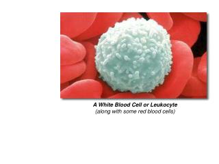
White Blood Cell Abnormalities. Laboratory Procedures. Let us recall what looks Normal. Some terminology for morphology. -penia : decreased number of cells in the blood (Neutropenia, lymphopenia). -philia or –cytosis : increased number of cells in the blood (neutrophilia, lymphocytosis).

E N D
White Blood Cell Abnormalities Laboratory Procedures
Some terminology for morphology • -penia: decreased number of cells in the blood (Neutropenia, lymphopenia). • -philia or –cytosis: increased number of cells in the blood (neutrophilia, lymphocytosis). • Macrocytosis: larger than normal cells • Microcytosis: smaller than normal cells • Anisocytosis: cells that are unequal in size • Left shift: presence of immature neutrophils in blood.
Nuclear Hyposegmentation • Can be found in cells that contain lobulated or segmented nuclei. • Which cells would this be? • What could this indicate?
Pelger-Huet Anomaly • Hyposegemented neutrophils that function normally. • Hereditary disorder; failure of the nucleus in mature cells to undergo segmentation.
Nuclear Hypersegmentation • Recall what can cause this.
Toxic Neutrophils • Characterized by ctyoplasm basophilia, Dohle bodies, toxic granulation, and/or foamy cytoplasm. • Cells have decreased functional abilities. • Animal with toxic, degenerative shift may be compromised by lack of adequate cell number and decrease ability of cells to function.
Dohle Bodies • Blue cytoplasmic inclusions. • Low numbers may be found in healthy cats. • Indicates toxicity in other species.
Intracytoplasmic Neutrophil Inclusions • Found in neutrophils of animals with certain infectious diseases. • Ehrlichia species.
Atypical or Reactive Lymphocytes • Contain azurophilic granules. • Generally associated with disease such as ehrlichiosis • May have cleaved nuclei • May have increased cytoplasm • May have increased basophilia in cytoplasm • Changes caused by antigenic stimulation secondary to vaccination or infection.
Lysosomal Storage Disorders • Rare • Inherited disease where substance is abnormally is stored within the cells due to enzyme deficiency. • Can cause skeletal or neurologic disorders • May contain vacuoles or certain granules.
Birman Cat Neutrophil Granulation Anomaly • Contain fine eosinophilic to magenta granules. • Inherited autosomal-recessive trait • Neutrophil function is normal and cats are healthy.
Chediak-Higashi Syndrome • Neutrophils in cats have large fused lysosomes within the cytoplasm. • Stain pink or eosinophilic. • May have tendency to bleed because platelet function is abnormal. • Generally are healthy cats.
Smudge Cells • May be called basket cells • Degenerative leukocytes that have ruptured. • Small numbers are not considered significant. • May be artifact when blood is held too long. • May be associated with leukemia.