Download
1 / 48
550 likes | 1.18k Views
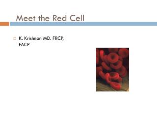
THE RED BLOOD CELL. Three aspects of red cell metabolism are crucial for normal erythrocyte survival and function: the red blood cell membrane haemoglobin structure and function active red cell metabolic pathways. A tissue section showing red cells in a small blood vessel.

E N D
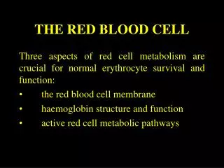
THE RED BLOOD CELL Three aspects of red cell metabolism are crucial for normal erythrocyte survival and function: • the red blood cell membrane • haemoglobin structure and function • active red cell metabolic pathways
A tissue section showing red cells in a small blood vessel. The red cell must be able to change shape and squeeze through small capillaries.
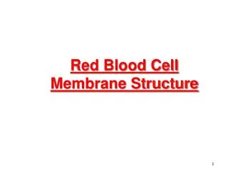
THE RED CELL MEMBRANE • The red cell membrane consists of a bipolar lipid layer supported by structural proteins. 50% of the membrane is protein 40% is lipid 10% is carbohydrate.
Structure of the red cell membrane. Spectrin Ankyrin Actin HAEMOGLOBIN INSIDE CELL Band 4.1 Lipid bilayer Cholesterol Fatty acid chain Phospholipid OUTSIDE CELL Band 3 Glycophorin PLASMA
Lipids consist of 60% phospholipid 30% neutral lipids (mainly cholesterol) 10% glycolipids. The phospho- and glycolipids are structural with polar groups (hydrophilic) on the external and internal surfaces of the cell. Non-polar groups (hydrophobic) form a barrier at the centre of the membrane.
The lipid bilayer forms the wall of the red cell and separates the contents from the external environment. Oxygen can pass through the lipid barrier and bind to the Hb inside. Alteration in lipid composition can produce target cells or acanthocytes.
Donor nucleotide (UDP-GalNAc) Acceptor sugar (Galactose) A gene product (A transferase) Gal GalNAc Glc Gal GlcNAc 1 band 3 1 4 4 2 3 2 3 Type 2 precursor Note: 14 linkage Red cell membrane Specific 13 linkage Fuc Carbohydrates are mostly found on the external surface of the red cell membrane. Monosaccharides are associated with specific blood group antigens, e.g. ABH and Lewis. Producing blood group A antigen on the red cell of a group A individual.
Proteins are either peripheral or integral. Integral proteins, e.g. glycophorin, are important for the active transport of solutes across the membrane. Spectrin, actin and ankyrin are peripheral proteins on the inner surface and maintain the biconcave shape of the red cell.
THE RED CELL "SKELETON" The lipid bilayer is stabilised by a protein framework on the inside of the cell. The "skeleton" is made of spectrin, an asymmetric two-chained molecule which is attached to the inside of the cell wall by other proteins including actin and ankyrin. The ankyrin binds to an integral protein (band 3) and the actin to a peripheral protein (band 4.1).
Spectrin gives the cell membrane its flexibility and strength. The red cell distorts as it passes through tiny capillaries, but once through the capillary, it immediately returns to its biconcave shape. If spectrin is denatured, e.g. by heat, the red cell assumes a spherical shape and loses its flexibility (spherocytosis).
PROTEIN IDENTIFICATION Proteins in the red cell membrane can be solubilised by a detergent called sodium dodecyl sulphate (SDS) and then be separated according to their size using polyacrylamide gel-electrophoresis (SDS-PAGE).
RED CELL ANTIGENS Blood group antigens are associated with the red cell membrane and are either integral to its structure or are adsorbed onto it from the plasma. They are made of proteins or carbohydrates. An individual has a particular antigen if the genes controlling its production are inherited. Protein antigens are under direct genetic control, whereas genetically controlled transferase enzymes assemble carbohydrate antigens.
HAEMOGLOBIN STRUCTURE Haemoglobin (MW 68,000) constitutes 95% of the red blood cell’s dry weight. 65% of haemoglobin synthesis occurs during the nucleated stages of RBC maturation and 35% occurs during the reticulocyte stage.
Normal Hb consists of globin (a tetramer of two pairs of polypeptide chains) and four haem groups. Each haem group contains a protoporphyrin ring plus ferrous iron (Fe++). The mitochondria are the main site of protoporphyrin synthesis. Iron is supplied from circulating transferrin and globin chains are synthesised on ribosomes.
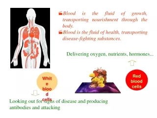
HAEMOGLOBIN SYNTHESIS The role of red cells is to carry oxygen to the tissues and to return carbon dioxide from the tissues to the lungs. Haemoglobin is essential for normal red cell function.
Six Hb variants are normally formed. Embryonic haemoglobins include Gower 1, Gower 2 and Hb Portland. HbF is the predominant haemoglobin of fetal life (65-95%). Adults have only trace amounts of HbF (<1%). HbA (>95%) and HbA2 (2.5-3.5%) are the main adult haemoglobins.
Globin chains found in adult haemoglobins are designated alpha (α) beta (β) gamma (γ) delta (δ). Hb A has two alpha and two beta chains (α2β2), Hb F has two alpha and two gamma chains (α2γ2) Hb A2 has two alpha and two delta chains (α2δ2). The alpha chain is thus common to all three types of adult Hb.
Alpha chain synthesis is directed by two α genes, α1 and α2, on chromosome 16. Beta and delta chains result from single genes on chromosome 11. The gamma chain is directed by two genes, G and A, on chromosome 11. Alpha chains have 141 amino acids and non-alpha chains have 146; the exact sequence of the amino acids has been determined.
HAEM consists of four pyrrole rings with a central iron atom linked to the four nitrogen atoms. The iron atom has two further binding sites, one of which is bound to a globin histidine residue and the other binds reversibly to oxygen.
Haem is synthesised mainly in mitochondria of erythroblasts, some steps occur in the cytoplasm. The initial and rate-limiting step is the fusion of succinyl-Co A with glycine mediated by ALA synthetase to form δ-aminolaevulinic acid (ALA). This occurs in the mitochondrion and depends on the presence of vitamin B6 (pyridoxal phosphate). The reaction is stimulated by erythropoietin and inhibited by haem.
The next step occurs in the cytoplasm. Two molecules of ALA fuse to form porphobilinogen. A double enzyme step forms uroporphyrinogen that is decarboxylated to coproporphyrinogen. At this point, the pathway re-enters the mitochondrium where protoporphyrin is formed.
Finally, ferrous iron is inserted to form haem by haem synthetase. The ferrous iron atom in each haem molecule is attached to the proximal histidine residue of a globin chain, but not to the distal histidine residue. Amino acids lying in the loop between proximal and distal histidine residues form the haem pocket, essential for the O2 carrying capacity.
HAEMOGLOBIN FUNCTION Red cells carry oxygen from the lungs to the tissues and return in venous blood with carbon dioxide. As the haemoglobin molecule loads and unloads O2, the individual globin chains in the haemoglobin molecule move in relation to each other. When O2 is unloaded, the β chains are pulled apart, permitting entry of the metabolite 2,3 diphospho-glycerate (2,3-DPG) resulting in a lower affinity for O2.
During oxygenation the two β chains move together to give a species more avid for O2. Hence as the initial oxygen is taken up by Hb it increases its affinity for oxygen to bind to the remaining haem groups in the molecule. The relationship between oxygen concentration in the blood (the partial pressure of oxygen) and the proportion of oxygen bound to haemoglobin (the percentage oxygen saturation) forms a sigmoid curve called the Hb oxygen-dissociation curve.
The P50O2 (the partial pressure of O2 at which Hb is half saturated) of normal blood is 27 mmHg. With increased affinity for O2, the curve shifts to the left (the P50 falls). With decreased affinity for O2, the curve shifts to the right (the P50 rises). Normally, O2 exchange operates between 95% saturation (arterial blood) with a mean arterial O2 tension of 95 mmHg and 70% saturation (venous blood) with a mean venous O2 tension of 40 mmHg.
The normal position of the curve depends on the concentration of 2,3-DPG, H+ ions and CO2 in the red cell and on the structure of the haemoglobin molecule. High concentrations of 2,3-DPG, H+ or CO2, and the presence of certain haemoglobins, e.g. HbS, shifts the curve to the right. This facilitates the release of oxygen from the red cells. HbF (unable to bind 2,3-DPG) and some rare abnormal haemoglobins associated with polycythaemia shift the curve to the left and so give up O2 less readily.
Mechanisms to Compensate for Anaemia A patient suffering from an anaemia caused by a loss of red cells may be able to compensate by shifting the oxygen dissociation curve to the right, making available red cells, though fewer in number, more efficient. A shift to the right may also occur in response to acidosis or a rise in body temperature.
A right shift of the oxygen dissociation curve is only one way in which patients may compensate for various types of hypoxia; other ways include increase in total cardiac output and in erythropoiesis.
RED BLOOD CELL METABOLISM Red cells generate energy almost exclusively through the anaerobic breakdown of glucose. The metabolism of the anucleated erythrocyte is more limited than that of other body cells. Mature red cells possesses little ability to metabolise fatty acids or amino acids and have no mitochondrial apparatus for oxidative metabolism. Red cells deliver oxygen, not consume it.
THE EMBDEN-MEYERHOF PATHWAY (EMP) In the Embden-Meyerhof Pathway (anaerobic) glucose is metabolised to lactate. For each molecule of glucose, two molecules of ATP are generated.
ATP provides energy for maintenance of red cell volume, shape and flexibility. The red cell has an osmotic pressure five times that of plasma. A membrane ATP-ase sodium pump ensures the correct concentrations of Na+ and K+ within the cell to protect the membrane from lysis. One molecule of ATP moves three sodium ions out and two potassium ions into the cell.
The Embden-Meyerhof pathway also generates NADH that is used to reduce non-functional methaemoglobin, containing ferric (Fe+++) iron, to functional reduced Hb, containing ferrous (Fe++) iron. 2,3-diphosphoglycerate (2,3-DPG) is generated by the Luebering-Rapoport shunt and forms a 1:1 complex with Hb to regulate haemoglobin's oxygen affinity.
THE PENTOSE PHOSPHATE PATHWAY (hexose monophosphate shunt) 5 to 10% of glycolysis occurs through the Pentose Phosphate Pathway (aerobic). Glucose 6-phosphate is converted to 6-phospho-gluconate and so to ribulose 5-phosphate. NADPH is generated and linked with glutathione to protect against oxidative stress e.g. hydrogen peroxide produced by oxidative drugs or phagocytes. NADPH is also used to maintain Hb in the active ferrous (Fe++) state.
One of the commonest inherited abnormalities of red cells is glucose-6-phosphate dehydrogenase (G6PD) deficiency in which the red cells are susceptible to oxidant stress.
Heinz Body Haemolytic Anaemia Heinz bodies G6PD deficiency is the most common enzymopathy causing hereditary haemolytic anaemia. Bite cells and helmet cells Blister cell
LABORATORY FINDINGS Features of increased erythrocyte breakdown: • Unconjugated bilirubinaemia. • Urobilinogenuria. • Haptoglobins decreased. • Radioisotope red cell survival studies can quantitate rate and site of destruction.
Features of increased erythrocyte production: • Reticulocytosis • Polychromasia and nucleated red cells in peripheral blood film. • Erythroid hyperplasia in bone marrow aspirate. • Radiological changes, e.g. "hair on end" appearance of cranial X-ray.
Features specific to intravascular haemolysis: • Haemoglobinaemia • (haptoglobin and haemopexin exhausted). • Methaemoglobinaemia. • Haemoglobinuria. • Haemosiderinuria.