
BACKGROUND
Universidad de Alcalá. Activation-Induced Cell Death As a Key Event on Peripheral Blood Mononuclear Cells from Allergic Patient. [Bautista Fortuna Patricia, Cerdán Sánchez Patricia, Gómez Serrano María, Del Monte Monge Alberto, Poquet Aparicio María Teresa, Prats Valcárcel Silvia ]**

BACKGROUND
E N D
Presentation Transcript
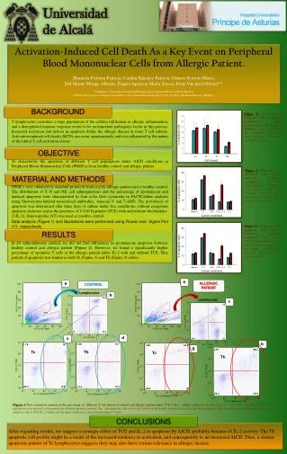
Universidad de Alcalá Activation-Induced Cell Death As a Key Event on Peripheral Blood Mononuclear Cells from Allergic Patient. [Bautista Fortuna Patricia, Cerdán Sánchez Patricia, Gómez Serrano María, Del Monte Monge Alberto, Poquet Aparicio María Teresa, Prats Valcárcel Silvia]** **Alumnos 5º Biología, Facultad de Biología de la Universidad de Alcalá de Henares (Edificio de Ciencias, Campus Universitario: Ctra. Madrid-Barcelona Km 33,600. E-28871 Alcalá de Henares,-Madrid-) BACKGROUND Figure 2. Percentage of apoptoticcells of differentperipheralbloodmononuclearcellsubsets in Medium. The vertical bluebarsrepresentthehealthy control data, and the red onestheallergicpatient. BACKGROUND T lymphocytes constitute a large population of the cellular infiltration in allergic inflammation, and a disregulated immune response seems to be an important pathogenic factor in this process. Increased activation and defects in apoptosis define the allergic disease in some T cell subsets. Activation-induced cell death (AICD) can occur spontaneously and it is influenced by the nature of the initial T-cell activation events. OBJECTIVE To characterize the apoptosis of different T cell populations under AICD conditions in Peripheral Blood Mononuclear Cells (PBMCs) from healthy control and allergic patient. Figure 3. Percentage of apoptoticcellsunderdifferentcultureconditions of Tc (CD3+CD8+) subset. The vertical bluebarsrepresentthehealthy control data, and the red onestheallergicpatient. MATERIAL AND METHODS PBMCs were obtained by standard protocols from a poly-allergic patient and a healthy control. The distribution of T, B and NK cell subpopulations and the percentage of spontaneous and induced apoptosis were characterized by four color flow cytometry in FACSCalibur analyzer using fluorocrome-labeled monoclonal antibodies, Annexin-V and 7-ADD. The prevalence of apoptosis was determined after three days of culture under five conditions: without exogenous apoptosis inducers and in the presence of T Cell Expander (TCE) with and without Interleukine-2 (IL-2). Staurosporine (ST) was used as positive control. Data analysis (Figure 1) and illustrations were performed using FlowJo and Sigma Plot v11, respectively. Figure 4. Percentage of apoptoticcellsunderdifferentcultureconditions of Th (CD3+CD8-) subset. The vertical bluebarsrepresentthehealthy control data, and the red onestheallergicpatient. RESULTS In all subpopulations studied, we did not find differences in spontaneous apoptosis between healthy control and allergic patient (Figure 2). However, we found a significantly higher percentage of apoptotic T cells in the allergic patient under IL-2 with and without TCE. This pattern of apoptosis was similar in both Tc(Figure 3) and Th(Figure 4) subset. e a ALLERGIC PATIENT CONTROL Lymphocytes b Lymphocytes f d c h g Tc Th Tc Th Figure.1Flow cytometric analysis of the percentage of different T cell subsets of control and allergic patient under TCE + IL-2 culture conditions. In both cases, Tc (CD3+CD8+) and Th (CD3+CD8-) populations were plotted to determinate the different apoptotic profiles. The total apoptotic cells was circled in blue in the healthy control and in red in the allergic patient. The comparation between these percentages of apoptotic cells in TCE+IL-2 culture and the other conditions is represented in figures 3 and 4. CONCLUSIONS After regarding results, we suggest a synergic effect of TCE and IL-2 in apoptosis by AICD, probably because of IL-2 activity. The Th apoptotic cell profile might be a result of the increased tendency to activation, and consequently to an increased AICD. Thus, a similar apoptosis pattern of Tc lymphocytes suggests they may also have certain relevance in allergic disease.


