VIRAL MYOCARDITIS
910 likes | 1.63k Views
VIRAL MYOCARDITIS. DR JULIAN JOHNY THOTTIAN DM CARDIO RESIDENT CALICUT MEDICAL COLLEGE. INTRODUCTION. Inflammation of heart muscle Most common agents- viruses, parasites and autoimmune state

VIRAL MYOCARDITIS
E N D
Presentation Transcript
VIRAL MYOCARDITIS DR JULIAN JOHNY THOTTIAN DM CARDIO RESIDENT CALICUT MEDICAL COLLEGE
INTRODUCTION Inflammation of heart muscle Most common agents- viruses, parasites and autoimmune state Pathology – Cardiac injury followed by immunologic response from host & agent resulting in cardiac inflammation leading to • Acute destruction • Persisting inflammation leading to cardiomyopathy >>> heart failure >>>death
Inflammation can be due to any reason like ischaemia , trauma , genetic but the classical type is due to exposure to discrete external antigens or internal triggers( auto immune) Incidence reported 8-10/ 100,000 population • Autopsy series is 1-5/100 Gore I, Saphir Am Heart J 1947,34:827-830 Pathologic series in young adults -8.6% Recent prospective postmortem data have implicated myocarditis in sudden cardiac death of young adults at rates of 8.6% to 12%.Fabre A, Sheppard MN. Sudden adult death syndrome and other non ischaemic causes of sudden cardiac death: a UK experience. Heart. 2005
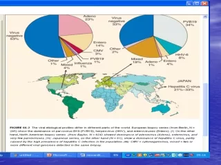
Epidemiology • Diagnosis of exclusion • Previously, the most common etiologic agents have been Enterovirus, with Coxsackie virus predominating. Dominance of Coxsackievirus has been replaced by a broader spectrum of viral causes, including Adenovirus, Parvovirus, and CMV
Kuhl and colleagues’ series • 245 patients with dilated cardiomyopathy were studied and etiology determined 51.4% - Parvovirus B19 21.6% - HHV 6 9.4% - Enterovirus 1.6% - Adenovirus 27.3%- Multiple infections
Bowles and coworkers series • 624 patients studied using PCR &Viral positivity in 38% • 22.8% - Adenovirus 13.6% -Enterovirus 1.0% - Parvovirus. Patient group was younger, resided mainly in North America
In Japan, Hepatitis C virus infection of the heart with HCM phenotype , dominated the etiologic profile Both Hepatitis C virus antibodies and genome have been detected in the serum and myocardial biopsy specimens of patients with myocarditis. Sato Y, Yamada T, Matsumori A Kluwer Academic publishers 2003: 325–339
Viruses • Coxsackie virus enter the system via CAR receptor • Adenovirus also uses the CAR receptor and Integrin receptor • Parvo virus causes endothelial dysfunction contributing to local inflammation and vasospasm.
Hepatitis C virus- mainly seen in Asian countries like Japan • Symptoms of myocarditis seen in 3rd week of illness • HIV -Postmortem analysis , 14 of 21 patients (67%) had criteria for myocarditis which increases to 83% in another study when one concentrates only on high-risk patients. Asymptomatic patients progression to DCM has been estimated to be around 15.9/1000. Ventricular dysfunction may be due to 1.HIV infection itself 2.Immunologic dysregulation 3.Side effects of antiretroviral treatment 4.Opportunistic coinfection or comorbid conditions
HIV-related myocarditis is lymphocytic myocarditis. The incidence was higher among patients with CD4+ counts <400 cells/mm3. Histological evidence of myocarditis was detected in 63 of 76 (83%) of these high-risk patients. FISH identified HIV-infected myocytes in 58 of 76 (76%) of this population.Barbaro G, Di Lorenzo G, Grisorio B, Barbarini G N Engl J Med. 1998; 339: 1093–1099.
Influenza- 5% -10% of infected patients. The presence of pre-existing cardiovascular disease greatly increases the risk of morbidity and mortality. Cardiac involvement typically occurs within 4 days to 2 weeks of the onset of the illness. • Mixed infections – Multiple viruses can increase each other virus` virulence. Seen in coxsackie and adenovirus infections
Pathophysiology of myocarditis Cooper LT Jr: Myocarditis. N Engl J Med 360:1526, 2009.
Immune activation and persistence • Knowlton and colleagues have identified that the enteroviral protease 2A cleaves the dystrophin-sarcoglycan complex located at the myocyte–extracellular matrix junction and leading to myocyte remodeling and subsequent cardiac dilation.
Innate immunity activation • The innate immunity is triggered by the foreign virus by the ubiquitous toll-like receptors (TLRs). • They recognize general molecular patterns TLR3 -dsRNA TLR4 -bacterial LPS. Activation leads to translocation of transcription factors such as NF-κB amplified cytokine production and interferon regulatory factors, leading to interferon production . The activation of TLRs signals also through adaptors and kinases such as MyD88 and interleukin receptor–associated kinases (IRAKs).
Downregulation of MyD88 and in turn of NF-κB and activation of acquired immunity are accompanied by the upregulation of type I interferons (IFN-α and IFN-β). Interferon is critical for host protection and survival, and its absence leads to excessive viral proliferation and direct cardiac damage
Cardiac remodelling • The virus can directly enter the endothelial cells and myocytes and interferes with the host protein synthesis and signaling pathways lead to direct cell death or hypertrophy. It can also modify the myocyte cytoskeleton and lead to dilated cardiomyopathy. Both innate and acquired immunity can lead to cytokine release and activation of MMPs that digest the interstitial collagen and elastin framework of the heart.
A retrospective study of 112 consecutive patients with biopsy-confirmed myocarditis at the Massachusetts General Hospital demonstrated the following pathological distribution: 1. Lymphocytic 55% 2. Borderline 22% 3. Granulomatous 10% 4. Giant cell 6% 5. Eosinophilic 6% Magnani JW, Suk-Danik HJ, Dec GW, DiSalvo TG Am Heart J. 2006
Clinical presentation • Presentation can range from asymptomatic state to symptoms of cardiac dysfunction, arrhythmias or heart failure, and hemodynamic collapse • Bimodal age distribution- a/c presentation in young while subtle & insidious often with cardiac failure with elderly.
Acute myocarditis • In a series -245 patients Fatigue- 82% DOE- 81% Arrhythmia -55% (SVT & VT) Palpitations -21% Chest pain -26% • Prodromal symptoms like fever, chills, myalgia -20-80% Kuhl U, Pauschinger M, Noutsias M Circulation 2005; 111:887
Fulminantmyocarditis • 10% of cases • Abrupt onset with 2weeks of illness • Hemodynamic compromise • Diffuse global hypokinesia and rarely cardiac dilation and typical thickening of ventricular wall. • On follow-up, 93% of the original cohort were alive and transplant free for 11 years after initial biopsy, compared with only 45% of those with chronic myocarditis
Giant cell myocarditis • More insidious and subtle • Presents with heart failure, arrhythmia, or heart block, which despite standard medical therapy fails to improve. The survival for this population is less than 6 months and is improved with the use of immunosuppressive therapy • Endomyocardial biopsy reveals a distinctive pattern of giant cells with active inflammation and scar tissue • Associated with Crohns disease and Thymoma
Chronic active myocarditis • Most older adults • Insidious and moderate ventricular dysfunction • Pathologic examination of a myocardial biopsy specimen may show active myocarditis, but more frequently it is only borderline or generalized chronic myopathic changes with fibrosis and myocyte dropout.
Diagnostic approaches • Lab investigations • ECG • 2D echo • CMR • Myocardial biopsy • Molecular evaluation
Lab investigations • CK or CK-MB is too insensitive –overall 8% • Troponins are more useful when high sensitivity thresholds are used. • Serum trop T >0.1ng/ml is used as cut off-sensitivity can be increased from 34% to 53% without compromising the specificity(94%), PPV-93% & NPV-56%. Lauer et al J Am CollCardiol. 1997; 30: 1354–1359. • Trop I – sensitivity 34% but specificity was 89% Smith et al Circulation. 1997; 95: 163–168 • Other biomarkers like cytokines, complements and antiviral or AHA are too insensitive or inadequately standardised.
ECG • T inversion-27% • ST elevation in 2 contiguous leads-54% ST depression-18% • Q waves in 18-27% • Bundle branch blocks • Prolongation of PR and QT intervals.Angelini A, Calzolari V Heart. 2000; 84: 245–250 • Kuhl and associates have found arrhythmias in 55% of patients including ventricular and supraventricular arrhythmias
ECHO 1. Ventricular dysfunction 2. Chamber dilation 3. Regional hypertrophy 4. RWMA Absence of regional coronary matching and rapid recovery of ventricular function gives clue to diagnosis Used as a follow up imaging modality. 42 patients with biopsy proven myocarditis identified Ventricular dysfunction in 69% of patients, but cardiac dilation is variable. RV dysfunction in 23%. RWMA in 64%. Reversible LV hypertrophy in 15% Pinamonti B, Alberti E Am J Cardiol. 1988; 62: 285–291.
2D ECHO helps to distinguish fulminant from more classic forms of myocarditis. Fulminantmyocarditis shows less diastolic dimensional increase but increased septal thickness, whereas the more classic forms of myocarditis show a much greater degree of ventricular dilation
Speckle tracking echocardiography: Potential diagnostic and prognostic tool in myocarditis In a retrospective study consisting of 45 patients with suspected acute myocarditis and 83 healthy controls who underwent this imaging procedure, event-free survival was significantly related to decline in longitudinal or circumferential strain and decline in longitudinal or circumferential strain rate, even among patients with preserved LV EF. Hsiao JF, Koshino Y, Bonnichsen CR, Yu Y, Miller FA Jr, Pellikka PA, et al. Speckle tracking echocardiography in acute myocarditis. Int J Cardiovasc Imaging. Feb 2013;29(2):275-84
MRI • Ability to characterize tissue according to water content and changes in contrast kinetics • Detects patchy disease Extracellular contrast agents such as gadolinium-DTPA will also distribute and clear very differently in inflamed or scarred tissue compared with normal tissue, leading to changes in T1 relaxation. Roditi et al studied 20 patients and found focal enhancement and RWMA in 18 of myocarditis patients using T1 wtd imaging and Gd Enhancement.
T2-weighted imaging strategy, such as the inversion recovery sequence helps in detection of myocarditic lesions showed a sensitivity of 84% and specificity of 74% based on biopsy or natural history evidence of myocarditis. Mahrholt et al studied gadolinium-enhanced MRI-guided biopsy of the right and left ventricles in 32 patients with suspected myocarditis. Left ventricular biopsy was generally performed from the region showing the most marked contrast enhancement. Biopsy of these specific myocardial regions resulted in PPV of 71% and NPV of 100%.
CMR suggested that the lateral wall may actually be the most common location for lesion development, not the septum. Mahrholdt H, Goedecke C, Wagner A, Meinhardt G, Athanasiadis A, Vogelsberg H, Firtz P, Klingel K, Kandolf R, Sechtem U. Circulation. 2004; 109: 1250–1258
Role of CMR in myocarditis( 2009) • (1) New-onset or persisting symptoms suggestive of myocarditis (2) Evidence of recent or ongoing myocardial injury or dysfunction (3) Suspected viral or nonischemic etiology.
The generally agreed CMR criteria of myocarditis include at least two (1) Regional or global myocardial signaling intensity increase in T2-weighted images (2) Increased global myocardial early gadolinium enhancement ratio between myocardium and skeletal muscle in gadolinium-enhanced T1-weighted images (3) At least one focal lesion with nonischemic regional distribution in inversion recovery prepared gadolinium-enhanced T1-weighted images (late gadolinium enhancement).
MYOCARDIAL BIOPSY • The Dallas criteria represented the first attempt to standardize the pathologic definition of myocarditis • The Dallas criteria require an inflammatory infiltrate and associated myocyte necrosis or damage not characteristic of an ischemic event • Chow and McManus first demonstrated insensitivity by biopsy. From a single endomyocardial biopsy sample, histologicmyocarditis could be demonstrated in only 25% of cases. Even with five random biopsy samples, correct diagnosis of myocarditis by the classic Dallas criteria could be reached in only about two thirds of subjects.
There are also variations in the interpretation of histologic samples by expert pathologists experienced in reading cardiac biopsies. 111 patients recruited in the original National Institutes of Health (NIH) Myocarditis Treatment Trial diagnosed with myocarditis by heart biopsy, only 64% had that diagnosis confirmed by the expert pathology panel during consensus reading of the same biopsy samples later.
Complication rates 2-5% • Almost half of these complications are vascular.
Molecular diagnostic techniques • In situ hybridization -Seeking the presence of viral genetic signatures in a pathologic sample. PCR amplification of the RNA from the biopsy specimen itself, have increased the sensitivity of detecting virus signatures in the heart. The presence of viral genome is entirely independent of the presence or absence of inflammatory cells on the same biopsy specimen. This shows that myocarditis is truly a disease of both the molecular trigger by the virus and the immunologic response by the host; either alone will be able to produce the disease syndrome
The tissues can also be analyzed for the upregulation of major histocompatibility complex (MHC) antigens. The sensitivity and specificity of MHC antigen upregulation have been shown to be 80% and 85%, respectively. MHC expression has been used to guide therapy for patients with myocarditis and inflammatory cardiomyopathy in one study evaluating immunosuppressive therapy. Wojnicz et al
Therapeutic approach • Supportive therapy • Immunosuppression • Interferon • Intravenous immunoglobulin • Immune adsorption • Immune modulation • Vaccination