Download
1 / 53
680 likes | 1.35k Views
Cardiomyopathy Myocarditis. Michelangelo L Sabas, MD FPCP FPCC FPSCCI Interventional Cardiology The Medical City Hospital Philippine Heart Center. Cardiomyopathy. group of diseases that primarily affect the heart muscle

E N D
CardiomyopathyMyocarditis Michelangelo L Sabas, MD FPCP FPCC FPSCCI Interventional Cardiology The Medical City Hospital Philippine Heart Center
Cardiomyopathy • group of diseases that primarily affect the heart muscle • not the result of congenital, acquired valvular, hypertensive, coronary arterial, or pericardial abnormalities
Two fundamental forms of cardiomyopathy • a primary type, consisting of heart muscle disease predominantly involving the myocardium and/or of unknown cause • a secondary type, consisting of myocardial disease of known cause or associated with a systemic disease such as amyloidosis or chronic alcohol use
Drawing comparing the morphologic classes of Cardiomyopathies.
Left and/or right ventricular enlargement; impaired systolic function, CHF, arrhythmias, emboli
CXR: moderate to marked cardiac silhouette enlargement, pulmonary venous hpn • ECG: ST segment and T wave abnormalities • Echo: LV dilatation and dysfunction • MPI: LV dilatation and dysfunction • Cath: LV dilatation and dysfunction, ↑ L and R filling pressures, ↓CO
Disproportionate LVH, with or without an intraventricular systolic pressure gradient; usually of a nondilated LV cavity
CXR: mild to moderate cardiac silhouette enlargement • ECG: ST segment and T wave abnormalities, LVH, abnormal Q waves • Echo: ASH, SAM of the MV • MPI: vigorous systolic function, perfusion defect • Cath: vigorous systolic function, dynamic LV outflow obstruction, ↑ L and R filling pressures
Endomyocardial scarring or myocardial infiltration resulting in restriction to left and/or right ventricular filling
CXR: mild cardiac silhouette enlargement • ECG: low voltage, conduction defects • Echo: ↑ LV wall thickness, normal or mildly reduced systolic function • MPI: normal or mildly reduced systolic function • Cath: normal or mildly reduced systolic function , ↑ L and R filling pressures
About one in three cases of CHF is due to dilated cardiomyopathy (DCM) • LV and/or right ventricular (RV) systolic pump function is impaired, leading to progressive cardiac dilatation (remodeling) • no cause is apparent in many cases • DCM is either familial or the end result of myocardial damage produced by a variety of known or unknown infectious, metabolic, or toxic agents • One-fifth to one-third of patients have familial forms of DCM
Clinical Manifestations • Chest pain • Syncope • Cardiac enlargement and findings of CHF • Pulse pressure is narrow and the ↑ JVP • S3 and S4 are common • MR and TR
Laboratory Examinations • CXR: enlargement of the cardiac • silhouette • ECG: ST or AF, ventricular arrhythmias, • LA abnormality, low voltage, diffuse • nonspecific ST-T-wave abnormalities, • and intraventricular and/or AV • conduction defects • Echo, CT imaging (CTI), and cardiac MRI show LV dilatation, with normal, • minimally thickened, or thinned walls, • and systolic dysfunction • BNP are usually elevated
Treatment • Standard therapy of heart failure with salt restriction • Anticoagulation • Antiarrhytmic agents – best avoided • Avoid: alcohol, NSAIDS, CCB’s • Resynchronization therapy • Implantable cardioverter- defibrillator (ICD) • Cardiac transplantation
Alcoholic CMP • Consumption of large quantities of alcohol (>80 grams of ethanol/day) • Partially genetically determined • ‘Holiday heart syndrome’
Peripartum CMP • Occurs during last trimester or within 6 months after delivery • Mortality: 10-20% • Avoid further pregnancies
Neuromuscular Disease • Duchenne’s progressive muscular dystrophy • Myotonic dystrophy • Friedreich’s ataxia
Drugs • Anthracycline derivatives, ie. Doxorubicin • Cyclophosphamide • Cocaine abuse
Arrhythmogenic Right Ventricular CMP / Dysplasia • Familial cardiomyopathy characterized by progressive fibrofatty replacement of the right ventricle and, to a much lesser degree, of the LV myocardium • RV failure with jugular venous distention, hepatomegaly, and edema • Ventricular tachyarrhythmias • ECG: QRS prolongation localized to the right precordial leads and left bundle branch block–type ventricular tachycardia • CTI and CMRI: RV dilatation, RV aneurysm, and fatty replacement
Taka-Tsubo CMP • Apical ballooning syndrome • Abrupt onset of severe chest discomfort preceded by a very stressful emotional or physical event • Women >50 years • ST-segment elevations and/or deep T-wave inversions in the precordial leads • No obstruction in the epicardial coronary arteries is noted on angiography • Reversible within 3–7 days and do not cause long-term cardiac dysfunction or disability
Features: • dynamic LV outflow tract pressure gradient • assymmetric LVH • Pathophysiologic abnormality: increased stiffness of the hypertrophied muscle (diastolic) • 50% of patients with HCM have a + family history = autosomal dominant
Clinical Features • Sudden death • Dyspnea • Angina pectoris, fatigue, syncope • PE: double or triple apical impulse, rapidly rising carotid arterial pulse, S4, systolic murmur
Hemodynamics • Pressure gradient is dynamic • Obstruction result from narrowing of the LV outflow tract by SAM of the MV against the hypertrophied septum • Basic mechanisms involved in the production and intensification of the dynamic intraventricular obstruction: (1) increased LV contractility, (2) ↓ventricular preload, and (3) ↓aortic impedance and pressure (afterload) • Hemodynamic features: ↑LVDP and systolic pressure gradient between the body of the LV and the subaortic region
Laboratory Evaluation • ECG: LVH, Q waves, Arrhythmias • Chest x-ray: normal or cardiomegaly • Echocardiogram: LVH, ‘ground-glass’ appearance of the septum, SAM of the MV • Apical hypertrophy: rare form of HCM
Treatment • Avoid: competitive sports / strenuous physical activity, dehydration, nifedipine • Caution on diuretics • Beta blockers for angina and syncope • Amiodarone • Verapamil, diltiazem • Pacemaker • Septal ablation, surgical myotomy /myectomy • Screen family members
Hallmark: abnormal diastolic function • Late stages: systolic function is also impaired. • Myocardial fibrosis, hypertrophy, or infiltration due to a variety of causes is responsible
Classification of Types of Restictive CMP Noninfiltrative Idiopathic cardiomyopathy Familial cardiomyopathy Hypertrophic cardiomyopathy Scleroderma Pseudoxanthoma elasticum Diabetic cardiomyopathy Infiltrative Amyloidosis Sarcoidosis Gaucher disease Hurler disease Fatty infiltration Storage Disease Hemochromatosis Fabry disease Glycogen storage disease Endomyocardial Endomyocardial fibrosis Hypereosinophilic syndrome Carcinoid heart disease Metastatic cancers Radiation Toxic effects of anthracycline Drugs causing fibrous endocarditis (serotonin, methysergide, ergotamine, mercurial agents, busulfan)
Clinical Features • Exercise intolerance and dyspnea are usually prominent • Dependent edema, ascites, and an enlarged, tender, and often pulsatile liver • Kussmaul's sign • Heart sounds may be distant, and S3 and S4
Laboratory • ECG: low-voltage, nonspecific ST-T-wave abnormalities and various arrhythmias • CXR: Absent pericardial calcification • Echo, CTI, and CMRI: symmetrically thickened LV walls and normal or slightly reduced ventricular volumes and systolic function; the atria are usually dilated • Doppler echo: diastolic dysfunction • Cath: ↓cardiac output, ↑ RV and LV end-diastolic pressures, and a dip-and-plateau configuration of the diastolic portion of the ventricular pressure pulses resembling constrictive pericarditis
Treatment • Management depends on the etiology and usually disappointing • anticoagulation
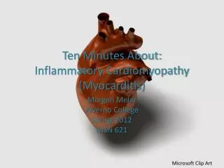
Myocarditis • cardiac inflammation due to infection, hypersensitivity to drugs, irradiation, chemicals or physical agents
Etiology • Viral (Most Common) Adenovirus Coxsackie virus/ Enterovirus Cytomegalovirus Parvovirus B19 Hepatitis C virus Influenza Human immunodeficiency virus Herpes virus Epstein-Barr virus Mixed infections • Bacterial Mycobacterial species Chlamydia pneumoniae Streptococcal species Mycoplasma pneumoniaeTreponema pallidum • Fungal Aspergillus Candida Coccidioides Cryptococcus Histoplasma
Etiology • ProtozoalTrypanosoma cruzi • Parasitic Schistosomiasis Larva migrans • Toxins Anthracyclines Cocaine • Hypersensitivity Clozapine Sulfonamides Cephalosporins Penicillins Tricyclic antidepressants • Autoimmune Activation Smallpox vaccination Giant cell myocarditis Churg-Strauss syndrome Sjögren syndrome Inflammatory bowel disease Celiac disease Sarcoidosis Systemic lupus erythematosus Takayasu arteritis Wegener granulomatosis
Clinical Features • history of a preceding upper respiratory febrile illness or a flulike syndrome, and viral nasopharyngitis or tonsillitis • transient electrocardiographic ST-T-wave abnormalities, to a fulminant condition with arrhythmias, acute CHF, and early death • simulates an acute coronary syndrome with chest pain, ECG changes, and elevated serum levels of troponin • physical examination is often normal