Download
1 / 25
270 likes | 556 Views
Myocarditis and pericarditis. Dr AliM Somily Prof Hanan A Habib. Introduction. Myocarditis is inflammatory disease of the heart muscle . Mild & self-limited with few symptoms or severe with progression to CHF & dilated CM Very localized or diffuse

E N D
Myocarditis and pericarditis Dr AliM Somily Prof Hanan A Habib
Introduction • Myocarditis is inflammatory disease of the heart muscle. • Mild & self-limited with few symptoms or severe with progression to CHF & dilated CM • Very localized or diffuse • Myocarditis can be due variety of infectious and non infectious causes • Viral infection is the most common cause • Others like toxin drugs and hypersensitivity immune.
Etiology, Epidemiology and Risk Factors • Epidemiology not accurate estimate of incidence as many cases are mild & brief and diagnosis is not made. • CoxsackievirusB is the most common cause of myocarditis • Other virus like coxsackievirus A, other echoviruses, adenoviruses influenza, EBV, rubella, vericella, mumps, rabies, hepatitis viruses and HIV. • Bacterial causes include corynebacteriumdiptheriae, syphilis Lyme disease or as a complication of bacterial endocarditis.
A parasitic cause includes toxoplasma gondii, chagas diseases, trichinellaspiralis, and Echinococcus. • Otherincludes rickettsia, fungi, Chlamydia, enteric pathogens, legionella and tuberculosis. • Giant cell myocarditisdue thymoma, SLE or thyrotoxicosis.
Clinical presentation • Days to weeks after onset of acute febrile illness or with heart failure without any known antecedent symptoms; highly variable • Fever, headache, muscle aches, diarrhea, sore throat and rashes similar to any viral infection • Chest pain, arrhythmias or sweating fatigue and may present with congestive heart failure.
Differential Diagnosis • Acute Myocarditis • Vasculitis • Cardiomyopathy ((drugs, radiation)
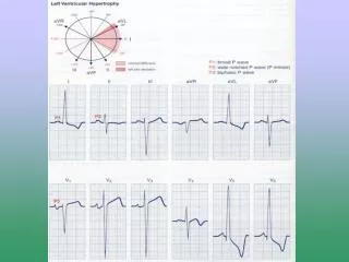
Diagnosis • WBCs, ESR, Troponins and CK-MB usually elevated • ECG (nonspecific ST-T changes and conduction delays are common) • Blood cultures • Viral serology and other specific test for Lyme, diphtheria and Chagasdisease maybe indicated on a case by case basis. • Chest X-rays show cardiomegaly • Radiology MRI and Echocardiogram • Heart muscle biopsy
Endomyocardial Bx • Pathologic exam may reveal lymphocytic inflammatory response with necrosis, but this is not sensitive b/c of the patchy areas of distribution. • “Dallas” criteria for histopathologic dx • May see “Giant cells”
Management • Often supportive; • Restricted physical activity in heart failure. • Specific antimicrobial therapy is indicated when an infecting agent is identified • Treatment of heart failure arrhythmia • Other drugs indicated in special situations like anticoagulant, NSAID steroid or immunosuppressive immunomodulatory agents. • Heart transplant
Management • Most cases of viral myocarditis are self limited. • One third of the patients are left with lifelong complications, ranging from mild conduction defects to severe heart failure. • Patient should be followed regularly every 1-3 months. • Sudden death may be the presentation of myocarditis in about 10% of cases.
Pathophysiology • Contiguous spread • lungs, pleura, mediastinal lymph nodes, myocardium, aorta, esophagus, liver • Hematogenous spread • septicemia, toxins, neoplasm, metabolic • Lymphangetic spread • Traumatic or irradiation
Pathophysiology • inflammation provokes a fibrinousexudate with or without serous effusion • the normal transparent and glistening pericardium is turned into a dull, opaque, and “sandy” sac • can cause pericardial scarring with adhesions and fibrosis
Pericarditis • Pericarditis is an inflammation of pericardium usually of infectious etiology • CoxsackievirusA and B, echovirus are the most common causes • Other includes herpes viruses, hepatitis B , mumps, influenza, adenovirus Varicellaand HIV
Bacterial Pericarditisusually complication of pulmonary infections (e.g. pneumonia empyema): S. pneumonia, M. tuberculosis, S. aureus, H. influenzae, K. pneumoniae legionella. • HIV patients may develop pericardial effusions (tuberculosis M. avium complex). • Disseminated fungal infection (Histoplasma, Coccidioides) • Parasitic infections (disseminated toxoplasmosis, contagious spread of Entamoebahistolytica)are rare causes.
Types of pericarditis • Caseouspericarditiscommonly tuberculosis in origin. • Serious Pericarditisby autoimmune diseases (rheumatoid arthritis, SLE). • Fibrous Pericarditis: A chronic Pericarditis usually caused by suppurative, caseous, or encased in a thick layer of scar tissue.
Types of Effusive Fluid • serous • transudative - heart failure • suppurative • pyogenic infection with cellular debris and large number of leukocytes • hemorrhagic • occurs with any type of pericarditis • especially with infections and malignancies • serosanguinous medslides.com
Constrictive Pericarditis • Idiopathic • radiotherapy • cardiac surgery • connective tissue disorders • dialysis • bacterial infection
Clinical presentation • Patients with Pericarditis will present with sudden pleuritic chest pain, fever, dyspnea and a friction rub. • Patient with tuberculouspericarditis has insidious onset of symptoms. • On examination exaggerated pulsusparadoxus JVP and tachycardia. • As the pericardial pressure increases, palpitations presyncope or syncope may occur.
Tuberculous Pericarditis • Incidence of pericarditis in patients with pulmonary TB ranged from 1-8% • Physical findings: fever, pericardial friction rub, hepatomegaly • Pericardial biopsy more definitive for diagnosis • TB skin test usually positive but no always specific • Fluid smear and culture for TB often negative medslides.com
Acute PericarditisDifferential Diagnosis • Acute myocardial infarction • Pulmonary embolism • Pneumonia • Aortic dissection
Diagnosis • ECG will show ST elevation, PR depression and T-wave inversion may occur later. • Blood culture • Leukocytosis and an elevated ESR are typical • Other routine testing urea and creatine. • PPD skin test is usually positive in tuberculousPericarditis. • Chest x-ray may show enlarged cardiac shadow or calcified pericardium and CT scan show pericardial thickening >5mm. • Pericardial fluid or pericardial biopsy specimens for fungi, antinuclear antibody tests and histoplasmosis complement fixation in endemic area.
Management • Management is a largely supportive for cases of idiopathic and viral Pericarditis including bed rest and NSAIDS, Colchicine. • Corticosteroid is controversial and anticoagulants usually contraindicated. • Specific antibiotics must include activity against S. aureus and respiratory bacteria. • Antiviral • Acyclovir for herpes simplex or varicellaganciclovir for CMV etc.
Management • Pericardiocentesis to relief tamponade. • Patients who recovered should be observed for recurrent. • Symptoms due to viral Pericarditis usually subsided within 1 month. • Uremic, rheumatic, collagen in 30% of patients include pericardial effusion and tamponade, constrictive Pericarditis and pleural effusion. • Restrictive Pericarditis and heart failure.