Download

1 / 16
170 likes | 646 Views
Viral Myocarditis. 附一儿科 林洁英. Introduction Myocarditis: Myocarditis is defined as inflammatory changes in the heart muscle and is characterized by myocyte necrosis .There are all kinds of causes and viral infections are the most common cause. Etiologies and Pathogenesy.

E N D
Viral Myocarditis 附一儿科 林洁英
Introduction • Myocarditis: Myocarditis is defined as inflammatory changes in the heart muscle and is characterized by myocyte necrosis .There areall kinds of causes and viral infections are the most common cause.
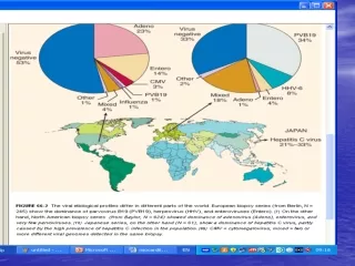
Etiologies and Pathogenesy • Virus infection: The most common causes of myocarditis are coxsackie A and B, echovirus,adenovirus and cytomegalovirus. • Directly damage • Auto-immunoreaction
Clinical manifestation • Symptoms: The presentation depends on the patient’s age and the acute or chronic process . fatigue, pale, chest malaise, myalgias, bellyache etc. Severe case: CHF, severe cardiac arrhythmia, cardiogenic shock, sudden death etc. • Signs: tachycardia, cardiac dilatation, heart sounds may be muffled and distant, gallop, signs of CHF and signs of cardiogenic shock.
Auxiliary Examination • Cardiac enzyme levels: CPK, LDH, CK-MB, cTnT、cTnI • Electrocardiogram: cardiac arrhythmia—premature beat, tachycardia, AV block, ST-T • Chest radiography: large cardiac sihouette, pulmonary venous congestion, edema
Auxiliary Examination • Echocardiography: cardiomegaly, ventricular disfunction, heart function↓ pericardial effusion ect. • Virology examination: viral isolation, specific antibody, PCR identification of a viral infection, myocardial biopsy etc.
Diagnosis Diagnosis may require blood tests, a chest X-ray, electrocardiogram or radionuclide angiocardiogram, and, in rare cases, biopsy of a tissue sample from the heart muscle.
Diagnosis • Clinical Findings 1. heart disfunction, cardiogenic shock or cardio-cerebral syndrome 2.cardiac dilatation ( chest radiography, echocardiography ) 3. change of electrocardiogram 4. CK-MB↑or cTnT(+)、cTnI(+)
诊断 心电图改变:以R波为主的2个或2个以上主要导联的ST-T改变持续4天以上伴动态变化;窦房、房室传导阻滞,完全右或左束支传导阻滞;成联律、多源、多型、成对或并行早搏;非房室结、房室折返引起的异位心动过速;低电压、异常Q波。
Diagnosis • Pathogenies 1. Diagnosis indexes: from endocardium, myocardial tissue, or pericardial fluid • Viral isolation • PCR identification of a viral infection • Specific antibody
Diagnosis • Pathogenies 2. Referenced diagnosis indexes • viral isolation from stool, blood etc • IgM(+) • PCR identification of a viral infection from blood
Diagnosis • Diagnosis guidelines 1.Clinical diagnosis of myocarditis: 2clinical findings 2. Diagnosis of viral myocarditis: 2clinical findings + 1 pathogenies of diagnosis indexes 3. Clinical diagnosis of viral myocarditis: 2clinical findings + 1 pathogenies of referenced diagnosis indexes
Diagnosis • Exclusion: rheumatism myocarditis toxic myocarditis congenital heart diseases cardiomyopathy endocardial fibroelastosis congenital AV block etc.
Stages • Acute phase: <6m • Deferable phase: 6m~1y • Chronic phase: >1y
Treatment • Rest: 3-4w, HD or HF 3-6m. • Protect cardiac muscle • Antioxidant: VitC, CoQ10, VitE • Antivirus • Corticosteroids and immunosuppression • Others