Download
1 / 67
670 likes | 684 Views
PK/PD approach for antibiotics: tissue or blood drug levels to predict antibiotic efficacy. PL Toutain National Veterinary school; Toulouse Wuhan 8 October 2015. Objectives of the presentation:. The three PK/PD indices Where are located the bugs ? Extracellular vs. intracellular

E N D
PK/PD approach for antibiotics:tissue or blood drug levels to predict antibiotic efficacy PL Toutain National Veterinary school; Toulouse Wuhan 8 October 2015
Objectives of the presentation: • The three PK/PD indices • Where are located the bugs ? • Extracellular vs. intracellular • Where is the biophase? • Interstitial space fluid vs. intracellular cytosol vs. intracellular organelles • How to assess the biophase antibiotic concentration • Total tissular concentration vs. ISF concentration. • The issue of lung penetration • Epithelial lining fluid (ELF):? • he hypothesis of targeted delivery of the active drug at the infection site by phagocytes • Plasma as the best surrogate of biophase concentration for PK/PD interpretation
First (scientific) consensus:The goal of PK/PD indices • The goal of PK/PD indices is to predict, in vivo, clinicaloutcomes: • Cure • prevention of resistance • Plasma free concentration is the relevant concentration for the establishment of a PKPD indice
Statements such as ‘concentrations in tissue x h after dosing are much higher than the MICs for common pathogens that cause disease’ are meaningless Mouton & al JAC 2007
For pulmonary infection, plasma free antibiotic concentration, not the epithelial Lining Fluid (ELF), is the best surrogate of biophase concentration
Second (marketing) consensus • It is more easy to promote a macrolide showing its high lung concentrations than its low plasma concentrations
MIC distribution for M haemolytica & P multocida (2004-20010) for tulathromycin
The PK/PD issue for macrolides (triamilides): plasma concentration lower than MICs • Good clinical efficacy and bacteriological cure with macrolides is achievable with plasma concentrations (much) lower, than the in vitro MICs for major lung pathogens MIC Cmax=0.5µg/mL ≤ to MIC90
MIC in MHB vs. calf serum25%,50%,75% and 100% MIC in MHB MIC in serum 100 % 50% 75% 25%
The case of tulathromycin • See presentation entitled “ how to establish a dosage regimen for a sustainable use of antibiotics
1: Where are located the pathogens and where is the biophase
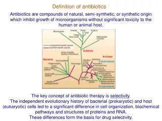
Where are located the pathogens ISF Most pathogens of clinical interest • S. Pneumoniae, E. Coli,Klebsiella • Mannhemia ; Pasteurella • Actinobacillius pleuropneumoniae • Mycoplasma hyopneumoniae • Bordetalla bronchiseptia Cell (most often in phagocytic cell) • Mycoplasma (some) • Chlamydiae • Brucella • Cryptosporidiosis • Listeria monocytogene • Salmonella • Mycobacteria • Rhodococcus equi
2: What are Antibiotic concentrations that are considered in the veterinary literature to explain antibiotic efficacy?
Antibiotic concentrations vs. efficacy • Total tissue concentrations • homogenates • biopsies • Extracellular fluids concentrations • implanted cages • implanted threads • wound fluid • blister fluid • ISF (Microdialysis, Ultrafiltration)
The inadequate tissue penetration hypothesis: Schentag 1990 • Two false assumptions • tissue is homogenous • bacteria are evenly distributed through tissue • spurious interpretation of all important tissue/serum ratios in predicting the antibacterial effect of AB Schentag, 1990
Total tissular concentration for betalactams and aminoglycosides • if a compound is distributed mainly extra-cellularly (betalactams and aminoglycosides), a total tissular concentration will underestimate the active concentration at the biophase by diluting the ISF with intracellular fluids.
Intracellular location of antibiotics Phagolysosome volume 1 to 5% of cell volume pH=5.0 Cytosol pH=7.4 Fluoroquinolones(x2-8) beta-lactams (x0.2-0.6) Rifampicin (x2) Aminoglycosides (slow Macrolides (x10-50) Aminoglycosides (x2-4) Ion trapping for weak base with high pKa value
Total tissular concentration for macrolides & quinolones • if a drug is accumulated in cells (the case for fluoroquinolones and macrolides), assays of total tissue levels will lead to gross overestimation of the extracellular biophase concentration.
4: what are the methods for studies of target site drug distribution in antimicrobial chemotherapy
PET images following administration of 18f-trovafloxacine Muller & al AAC 2004
Methods considered of limited interest for studies of target site drug distribution • Tools developed to determine antibiotic concentrations in various surrogates for the ISF and having no pathophysiologic counterpart in humans . • in vitro models, • fibrin clots, • tissue chambers, • skin chambers(blister) • wound exudates, • surface fluids, • implanted fibrin clots, • peripheral lymph. Muller & al AAC 2004
The tissue cage model for in vivo and ex vivo investigations
The tissue cage model • Perforated hollow devices • Subcutaneous implantation • development of a highly vascularized tissue • fill up with a fluid with half protein content of serum (delay 8 weeks) • C.R. Clarke. J. Vet. Pharmacol. Ther. 1989, 12: 349-368
PK in tissue cagein situ administration • PK determined by the cage geometry (SA/V ratio is the major determinant of peak and trough drug level) • T1/2 varies with the surface area / volume ratio of the tissue cage • Penicillin 5 to 20 h • Danofloxacin 3 to 30 h Greko, 2003, PhD Thesis
The Tissue cage model: veterinary application • To describe PK at site of infection (calves, dogs, horses…): NO • To assess the influence pf inflammation by comparing exudate and transudate concentration • To investigate PK/PD relationship: YES • ex vivo : killing curves (exudate/transudate) • in vivo : Greko (inoculation of the tissue cage)
What is microdialysis (MD)? • Microdialysis, a tool to monitors free antibiotic concentrations in the fluid which directly surrounds the infective agent
Microdialysis: The Principle • The MD Probe mimics a "blood capillary". • Diffusion of drugs is across a semipermeable membrane at the tip of an MD probe implanted into the ISF of the tissue of interest. • There is an exchange of substances via extracellular fluid
Microdialysis : Limits • MD need to be calibrated • Retrodialysis method • tedious. • The in vivo percent recovery is calculated (CV of about 10-20%)
MD need to be calibrated: A small experimental error in recovery estimate results in a relatively larger error in drug concentration estimates which is probably responsible for the greater interanimal variability observed in lung tissue than in the other media Marchand & al AAC June 2005
Ultrafiltration • Excessive (in vivo) calibration procedures are required for accurate monitoring • Unlike MD, UF-sample concentrations are independent on probe diffusion characteristics
Microdialysis vs. Ultrafiltration Microdialysis: a fluid is pumped through a membrane; Ultrafiltration Vacuum The driving force is a pressure differential (a vacuum) applied across the semipermeable membrane The analyte cross the membrane by diffusion The driving force is a concentration gradient
Marbofloxacin : plasma vs.ISFIn vivo filtration Microdialysis • Not suitable for long term in vivo studies Ultrafiltration • Suitable for long term sampling (in larger animals, the UF permits complete freedom of movement by using vacutainer collection method) Bidgood & Papich JVPT 2005 28 329
This study’s objectives were to determine intestinal antimicrobial concentrations in calves administered enrofloxacin or ceftiofur sodium subcutaneously, and their impact on representative enteric bacteria Ultrafiltration devices were implanted in the ileum and colon of 12 steers,
Enrofloxacin (SQ, 7.5mg/kg) • AUC (enro+cipro) • Plasma=19 (total) • ISF=25 (free) • Ileaum=21 (free) • Spiral colon =36 (free)
Ceftiofur SQ (2.2mg/kg) • AUC • Plasma=137 (total) • ISF=15 (free) • Ileum=40 (free) • Spiral Colon =34 free)
6-What we learnt with animal and human microdialysis studies
Total (plasma, muscle) free (plasma) interstitial muscle interstitial adipose tissue Plasma (total, free) concentration vs interstitial concentration (muscle, adipose tissue) (Moxifloxacin) 1000 Concentration (ng/mL) 100 2 6 10 12 40 20 30 Time (h) Muller AAC, 1999
Plasma (total, free) concentration vs muscle (free) concentration cefpodoxine Total (plasma) free (muscle) free (plasma) cefixime Liu J.A.C. 2002
What we learnt with animal and human MD studies • MD studies showed that: • the concentrations in ISF of selected antibiotics correspond to unbound concentrations in plasma and are much lower than concentrations reported from whole-tissue biopsy specimens. • Concentrations of beta –lactams and aminoglycosides in ISF are mostly in the range of free concentrations in serum • Concentration of quinolones and macrolides at their target site are considerably lower than those predicted from tissue biopsy specimens
What we learnt with animal and human microdialysis studies • Free plasma concentration is a good surrogate of most interstial fluid (ISF) concentration
MIC measured in MHB is homogeneous to a “free concentration”
Effect of protein binding on antimicrobial activity MICs of Staphylococcus aureus (Data from Kunin et al 1973) MIC (µg/mL) fb 0.22 0.65 0.90 0.95 0.37 0.93
The free concentration paradigm in pharmacokinetics is supported by MD findings Tissular space Blood/Plasma Interstitial fluid Tissue bound Plasma bound ISF bound Total BUG Measured by analytical technique Free Free Free Elimination
Tissue concentrations of levofloxacin in inflamed and healthy subcutaneous adipose tissue Hypothesis: Accumulation of fibrin and other proteins, oedema, changed pH and altered capillary permeability may result in local penetration barriers for drugs Methods: Free Concentrations measured by microdialysis after administration of a single intravenous dose of 500 mg. Inflammation No inflammation Results:The penetration of levofloxacin into tissue appears to be unaffected by local inflammation. Same results obtained with others quinolones Bellmann & al Br J Clin Pharmacol 2004 57
What we learnt with MD studies: Inflammation • Acute inflammatory events seem to have little influence on tissue penetration. • “These observations are in clear contrast to reports on the increase in the target site availability of antibiotics by macrophage drug uptake and the preferential release of antibiotics at the target site a concept which is also used as a marketing strategy by the drug industry”Muller & al AAC May 2004
Animal and human studies MD: The issue of lung penetration • Lung MD require maintenance under anesthesia, thoracotomy (patient undergoing lung surgery).. • Does the unbound concentrations in muscle that are relatively accessible constitute reasonable predictors of the unbound concentrations in lung tissue (and other tissues)?