Download
1 / 62
630 likes | 915 Views
Lateralization & The Split Brain and Cortical Localization of Language. Ch. 16. Outline. The Dominant Left Hemisphere Tests of Cerebral Lateralization The Split-Brain Experiment Tests of Split-Brain Patients Differences Between the Left and Right Hemispheres Broca’s Area Wernicke’s Area.

E N D
Lateralization & The Split Brain andCortical Localization of Language Ch. 16
Outline • The Dominant Left Hemisphere • Tests of Cerebral Lateralization • The Split-Brain Experiment • Tests of Split-Brain Patients • Differences Between the Left and Right Hemispheres • Broca’s Area • Wernicke’s Area
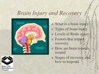
Aphasia and Apraxia:The Dominant Left Hemisphere • In 1836, Dax reported that not one of his 40 or so patients with speech problems had displayed damage restricted to the right hemisphere • 25 yrs later, Broca reported the results of the postmortem examination of two aphasic patients (patients with deficits in the use of language that are not attributable to general sensory, motor, or intellectual dysfunction)…
Aphasia and Apraxia:The Dominant Left Hemisphere • Both had diffuse left hemisphere damage that seemed to be centered in an area of the inferior left prefrontal lobe, just in front of the primary motor face area • This became known as Broca’s area that is associated with grammar and speech production
Aphasia and Apraxia:The Dominant Left Hemisphere • Liepmann discovered that apraxia (difficulty performing movements with either side of the body when asked to do so, but not when performing them spontaneously) was almost always associated with left-hemisphere damage
Aphasia and Apraxia:The Dominant Left Hemisphere • This led to the view that all complex activities were performed by the left hemisphere; the left and right hemispheres thus became known as dominant and minor hemispheres, respectively
Tests of Cerebral Lateralization • The first evidence of language laterality came from comparisons of the effects of left and right unilateral lesions; today, the sodium amytal test and dichotic listening test are commonly used to assess language laterality
Tests of Cerebral Lateralization • PET of FMRI techniques have revealed that there is typically more activity in the left hemisphere than the right during language-related activities
Tests of Cerebral Lateralization • Many studies have reported a relation between speech laterality and handedness; the following general conclusions have been reached:
Tests of Cerebral Lateralization • Nearly all (about 95%) right-handed subjects are left-hemisphere dominant for speech; • most left-handed or ambidextrous subjects (about 70%) are also left-hemisphere dominant for speech; and • Early left-hemisphere damage can cause the right hemisphere to become dominant for speech and the left hand to be preferred
The Split-Brain Experiment • In 1953, Myers and Sperry performed an experiment on cats that changed the way that we think about the brain; and it provided a means of comparing the function of the two hemispheres • It was designed to reveal the function of the brain’s largest commissure, the corpus callosum
The Split-Brain Experiment • Earlier studies failed to reveal any deficits in laboratory animals following callosal transection, and people born without a corpus callosum had been reported to be perfectly normal
The Split-Brain Experiment • In the Myers and Sperry experiment there were four groups of cats: • Corpus callosum severed • Optic chaims severed • corpus callosum and optic chiasm severed • Intact controls
The Split-Brain Experiment • In phase 1 of the experiment, all cats learned a lever-press pattern discrimination task with a patch over one eye; all four groups readily learned this simple task • In phase 2, the patch was switched to the other eye…
The Split-Brain Experiment • The cats in the optic-chiasm-severed group, corpus-callosum-severed group, and control kept performance same • In contrast the optic-chiasm-and-corpus-callosum-severed group acted as if the task were completely new to them - they had to learn it again with no savings
The Split-Brain Experiment • We can conclude: • The cat forebrain has the capacity to act as two separate forebrains, each capable of independent learning and of storing its own memories; • The function of the corpus callosum is to carry information between hemispheres • The best strategy for studying corpus callosum function is to use a method to limit information to a single hemisphere
Tests of Split-Brain Patients • Commissurotomy is performed on patients with life-threatening cases of epilepsy to reduce the severity of convulsions by restricting epileptic discharges to half of the brain
Tests of Split-Brain Patients • The operation is remarkably effective; many commissurotomized epileptic patients never experience another major convulsion; more remarkably they experience few obvious side effects in their daily lives
Tests of Split-Brain Patients • The controlled neuropsychological testing of these split-brain patients has revealed some amazing things about the human brain • To test split brain patients,visual stimuli are flashed to the right or left of a fixation point on a screen • Also tactual information is presented to one hand under a ledge or in a bag
Tests of Split-Brain Patients • These tests confirmed the conclusion that commissurotomized patients have two independent streams of consciousness
Evidence of Two Independent Streams of Consciousness • When an object was presented to the left hemisphere, either by touching something with the right hand or viewing something in the right visual field, the subject could:
Evidence of Two Independent Streams of Consciousness • Pick out the correct object with the right hand • Could not pick out the correct object with the left hand • Could name the correct object
Evidence of Two Independent Streams of Consciousness • When an object was presented to the right hemisphere, either by touching something with the left hand or viewing something in the left visual field, the subject could:
Evidence of Two Independent Streams of Consciousness • Could pick out the correct object with the left hand • Could not pick out the correct object with the right hand • Claimed nothing had been presented
Cross-cuing • Represents communication between hemispheres via a nonneural route • For example: a red or green light is flashed in the left visual field; the split-brain patient was then asked to name the color: red or green…
Cross-cuing • Most split-brain patients get 50% correct on this task (guessing, by chance); however one patient performed almost perfectly • When the performance of this subject was carefully monitored, it was noticed that on the trials when the patient initially said (left hemisphere) the incorrect color, his head shook and the patient then changed their guess to the other color
Cross-cuing • Apparently, the right-hemisphere (who knew the correct answer) heard the incorrect guess of the left hemisphere, and signaled to the left hemisphere that it was wrong by shaking the person’s head; when only first guesses were counted, performance fell to 50%
Learning Two Things at Once • Split-brain patients are capable of learning two things at once • If a split-brain patient is visually presented two objects at the same time - let’s say a pencil in the LVF and apple in the RVF - s/he can reach into two different bags at the same time, one with each hand, and pull out the two objects - a pencil in the left-hand and apple in the right
Helping-Hand Phenomenon • Occurs when the two hemispheres are presented with different information about the correct choice and then are asked to reach out and pick up the correct object from a collection in full view • Usually the right hand will reach out to pick out what the left hemisphere saw, but the right hemisphere seeing what it thinks is an error being made causes the left hand to grab the right hand and pull it over to the other object
Differences BetweenThe Left and Right Hemispheres • Language is the most lateralized of all abilities; the left-hemisphere is better than the right at most language-related tasks • however, the right hemisphere proved to be able to understand single written and spoken words; also right-hemisphere detects prosody and discourse
Differences BetweenThe Left and Right Hemispheres • The right hemisphere proved better than the left at a variety of tasks involving spatial ability, emotional stimuli and musical tasks
Differences BetweenThe Left and Right Hemispheres • The two hemispheres seem to engage different types of memory processing; LH attempts to place its experience in a larger context (relation of parts that make up the whole), while the RH attends strictly to the Gestalt perceptual characteristics of the stimulus (parts or whole but not relation between) • This is usually termed analytical (LH) versus holistic (RH)
Differences BetweenThe Left and Right Hemispheres • Thus the RH should not be regarded as the minor hemisphere; it has different abilities, not less important ones
Differences BetweenThe Left and Right Hemispheres • There are also anatomical asymmetries in the human brain; for example the planum temporale and frontal operculum (language related areas) are larger in LH • However, Heschl’s gyrus (also language related) in larger in RH
Differences BetweenThe Left and Right Hemispheres • (not in book) left-handers seem to have symmetrical planum temporales, suffer less severely from LH aphasia, and suffer more severely from RH aphasia • This suggests left-handers may have a more diffuse representation of language and is evident in differential grammatical strategies in sentence processing
Three Theories ofCerebral Asymmetry • Analytic-synthetic theory • Motor theory • Linguistic theory
Analytic-Synthetic Theory • Suggests that there are two fundamentally different modes of thinking, an analytic mode (LH) and synthetic mode (RH), and that the neural circuitry for each is fundamentally different • LH (pieces of the whole) operates in logical, sequential, analytic fashion • RH (the whole) makes immediate, overall synthetic judgments
Motor Theory • Posits that LH is specialized for fine motor movement of which speech is but one example • Two lines of evidence: • Lesions of the LH disrupt facial movements more than do RH lesions, even when they are not related to speech • Degree of disruption of nonverbal facial movements is positively correlated with the degree of aphasia
Linguistic Theory • Based on the view that the primary function of the LH is language; this is based on studies of deaf people who communicate using ASL; this ability is lost if these people suffer damage to the LH, even when they are able to make the movements required • (or is this just showing ASL is a language, and that language is highly analytical?)
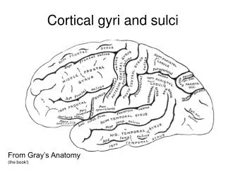
Broca’s Area • Inferior left prefrontal lobe in left hemisphere • Damage leads to deficits primarily speech production (problems with expression) and also grammatical comprehension
Wernicke’s Area • Left temporal lobe, just posterior to the primary auditory cortex • Damage leads to deficits to semantic language comprehension (problems with reception) and speech is imcomprehensible, despite having correct grammar, rhythm an intonation (word salad)
Conduction Aphasia • Damage to pathway connecting Broca’s and Wernicke’s areas called the arcuate fasciculus • Comprehension and spontaneous speech are intact but patient not able to repeat words they have just heard
Alexia • Damage to the left angular gyrus (area of left temporal and parietal cortex jost posterior to Wernicke’s) • Inability to read despite intact language comprehension and production
Agraphia • Also due to damage to the left angular gyrus • Inability to write despite intact language comprehension and production • Involvement of LAG in alexia and agraphia show its responsible for language related visual input
Wernicke-Geshwind Model • Seven components in Left hemisphere: primary visual cortex, angular gyrus, primary auditory cortex, Wernicke’s area, arucate fasciculus, Broca’s area, and primary motor cortex
Responding to a heard question • Primary auditory cortex to Wernicke’s area where comprehended • To respond, concept generated in Wernicke’s area, goes via arcuate fasciculus to Broca’s area, then to primary motor cortex and articulatory areas (face, lip, and tongue muscles, voice box, and muscles assoicated with lungs)
Reading aloud • Primary visual cortex to left angular gyrus, which transmits visual code to auditory code • Then to Wernicke’s area to arcuate fasciculus to Broca’s to primary motor cortex to articulatory areas
Evidence against W-G Model • Damage to these boundaries has little lasting effect on language • Damage to other brain areas can produce aphasia • Broca’s and Wernicke’s aphasia are rarely “pure” - aphasia is both receptive and expressive • Major individual differences for cortical localization for language