Download

1 / 8
80 likes | 200 Views
This comprehensive guide explores the posterior aspect of the forearm in 7 critical questions. It details the two muscle layers: the superficial and deep layers, including the specific muscles found in each. The guide also explains the movements these muscles facilitate, such as hand extension and finger abduction, and their innervation by the radial and posterior interosseous nerves. Additionally, it discusses the vascular supply from the radial and posterior interosseous arteries, providing essential anatomical insights for students and professionals alike.

E N D
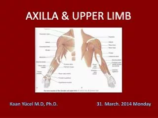
Posterior aspect of the forearm • IN 7 QUESTIONS • Kaan Yücel M.D., Ph.D • 28.December.2012 Friday
1. How are the muscles in the posterior compartment estabslihed? In two layers: a superficial layer a deep layer.
2. Which muscles are in the superficial layer? Lateral epicondyle of humerus Lateral epicondyle of humerus Brachioradialis Extensor carpi radialis longus Proximal part of supraepicondylar ridge of humerus Distal part of supraepicondylar ridge of humerus Extensor carpi radialis brevis Lateral epicondyle of humerus common extensor origin Lateral epicondyle of humerus common extensor origin Lateral epicondyle of humerus Extensor carpi ulnaris Extensor expansion (hood) of the 5th digit Extensor digitorum Olecranon and proximal posterior surface of ulna Extensor digiti minimi dorsal aspects of the bases of the middle and distal phalanges of the medial four digits Anconeus Lateral surface of distal end of radius proximal to styloid process Common originlateralepicondyle of the humerus Except for the brachioradialis and anconeus, extend as tendons into the hand. Base of 3rd metacarpal Base of 5th metacarpal Base of 2nd metacarpal Base of 2nd metacarpal Base of the 5th metacarpal Base of 3rdmetacarpals
Posterior surface of distal 1/3 of ulna and interosseous membrane Posterior surface of proximal halves of ulna, radius, and interosseous membrane 3. Which muscles are in the deep layer? Superficial (humeroulnar) head •lateral epicondyle of humerus •radial collateral and anular ligaments Deep (ulnar) head Supinator crest of the ulna Supinator Abductor pollicis longus Extensor pollicis longus Extensor pollicis brevis Base of 1st metacarpal Extensor indicis Posterior surface of middle third of ulna and interosseous membrane Dorsal surface of base of distal phalanx of thumb • Lateral surface of radius superior to the anterior oblique line Extensor expansion (hood) of 2nd digit Posterior surface of distal third of radius and interosseous membrane Dorsal surface of base of proximal phalanx of the thumb Except for the supinator muscle, all these deep layer muscles originate from the posterior surfaces of the radius, ulna, and interosseous membrane and pass into the thumb and fingers.
4. What movements do these muscles do? EXTENSION OF THE HAND & FINGERS ABDUCTION OF THE FINGERS SUPINATION
5. How are the muscles of the posterior compartment of the forearm innervated? Brachioradialis Extensorcarpiradialislongus Radialnerve (Main nerve) Supinator Extensor carpi radialis brevis Deep branch of radial nerve Rest Posterior interosseous nerve continuation of deep branch of radial nerve
6. ....arteries in the posterior compartment of the forearm? Radial artery Posterior interosseous artery origin: common interosseous branch of the ulnar artery recurrent interosseous artery End by joining to dorsal carpal arch of the wrist Anterior interosseous artery origin: common interosseous branch of the ulnar artery
7. ...radial nerve in the posterior compartment of forearm? Deep branch becomes posterior interosseous nerve after emerging from between 2 heads of supinator Posterior interosseous nerve passes deep to extensor pollicis longus to reach the wrist.