Download
1 / 57
570 likes | 680 Views
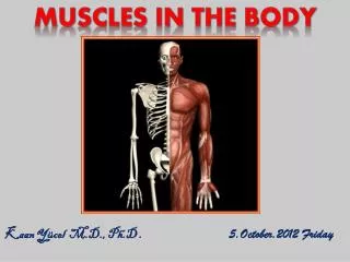
MUSCLES IN THE BODY. Kaan Yücel M.D., Ph.D . 5.October.2012 Friday. The muscular system consists of all the muscles of the body. The discipline related to the study of muscles is myology .

E N D
MUSCLES IN THE BODY Kaan Yücel M.D., Ph.D . 5.October.2012 Friday
The muscular system consists of all the muscles of the body. • The disciplinerelated to the study of muscles is myology. • Musculus (muscle) is derived from the word mus-mouse; musculus- little mouse. • So called because the shape and movement of some muscles (notably biceps) were thought to resemble mice. • If you bend and straighten your arm at the elbow, you should see the front of the upper arm move under the skin. To the ancient Romans this movement resembled a little mouse scurrying beneath the skin.
All skeletal muscles are composed of one specific type of muscle tissue. • These muscles move the skeleton, therefore, move the body parts.
Types of Muscles • based on distinct characteristics • Functional • voluntary vs. involuntary • Histological • striated vs. smooth or unstriated • Anatomical (location) • @ body wall (soma) and limbs • @ hollow organs (viscera) or blood vessels
Skeletal striated muscle • voluntarysomatic muscle • gross skeletal muscles that compose the muscular system • moving or stabilizing bones and other structures (e.g., the eyeballs). • Innervatedby the somatic nervous system.
Cardiac striated muscle • involuntary visceral muscle • forms the walls of the heart and adjacent parts of the great vessels. • pumps blood.
Smooth muscle (unstriated muscle) • involuntary visceral muscle • forms part of the walls of most vessels and hollow organs (viscera) • moving substances through them • coordinated sequential contractions (pulsations or peristaltic contractions). • Innervatedby the autonomic nervous system.
FEATURES OF SKELETAL MUSCLES HEAD OR BELLY fleshy, reddish, contractile portions TENDONwhite non-contractile portions composed mainly of organized collagen bundles, that provide a means of attachment.
Mostskeletalmusclesattachto • Directlyorindirectlytobones • Cartilages • Ligaments • Fascias • orcombinations of theonesabove Sometoorgans (eyeball)/skin (facialmuscles)/mucousmembranes(intrinsictonguemuscles)
Muscles are organs of locomotion (movement) also: provide static support give form to the body provide heat
Sometendons form flatsheetsaponeuroses anchor the muscle to the skeleton to deep fascia toaponeurosisof another muscle
Many terms provide information about a structure's • Shape • Size • Location • Function • Resemblanceof one structure to another
Basisof function Bones attachedto Abductordigitiminimimuscleabducts the little finger. Sternocleidomastoidmuscle (G. kleidos, bolt or bar, clavicle) attaches inferiorly to the sternum and clavicle and superiorly to the mastoid process of the temporal bone of the cranium. Levatorscapulae elevates the scapula (L. shoulder blade).
Descriptive names Deltoid muscle triangular, like the symbol for delta, the fourth letter of the Greek alphabet. -oid“like”; deltoid means like delta.
Position • medial, lateral, anterior, posterior • Length • brevis, short; longus, long • Shape • piriformismuscle • pear shaped (L. pirum, pear + L. forma, shape or form).
Location • temporalismuscle • in the temporal region (temple) of the cranium (skull).
CLASSIFICATION OF MUSCLES accordingtotheirshapes Flat muscles parallel fibers often with an aponeurosis External obliquemuscle broad flat muscle Sartorius narrow flat muscle with parallel fibers longestmuscle in the body
feather-like (L. pennatus, feather), arrangement of fasicles Unipennate Extensor digitorum longus Bipennate Rectus femoris Pennate muscles Multi-pennate Deltoid
spindle shaped with a round, thick belly (or bellies) and tapered ends Fusiformmuscles
Convergent muscles • arise from a broad area • converge to form a single tendon • four equal sides (L. quadratus, square) • rectus abdominis • between its tendinous intersections. Quadratemuscles
Circular or sphincteral muscles surround a body opening or orifice, constricting it when contracted orbicularis oculi closes the eyelids
Multi-headed or multi-bellied muscles more than one head of attachment or more than one contractile belly Biceps muscles two heads of attachment triceps muscles three heads Twobellies digastric muscle gastrocnemius muscle
Contraction of muscles Skeletal muscles function by contracting they pull and never push. When a muscle contracts and shortens one of its attachments usually remains fixed the other attachment(more mobile) pulled toward it movement
Attachments of muscles • origin&insertion • Originproximal end of the muscle • remains fixed during muscular contraction. • Insertiondistal end of the muscle • movable This is not always the case. Some muscles can act in both directions under different circumstances.
Whereas the structural unit of a muscle is a skeletal striated muscle fiber, the functional unit of a muscle is a motor unit, consisting of a motor neuron and the muscle fibers it controls.
When a motor neuron in the spinal cord is stimulated, it initiates an impulse that causes all the muscle fibers supplied by that motor unit to contract simultaneously.
The number of muscle fibers in a motor unit varies from one to several hundred. The number of fibers varies according to the size and function of the muscle. • Large motor units, in which one neuron supplies several hundred muscle fibers, are in the large trunk and thigh muscles. • Movement (phasic contraction) results from the activation of an increasing number of motor units, above the level required to maintain muscle tone.
Functions of muscles Prime mover (agonist) main muscle responsible for producing a specific movement of the body. Doesmost of the work (expending most of the energy) required. In most movements, there is a single prime mover, but some movements involve two prime movers working in equal measure.
Fixator steadies the proximal parts of a limb through isometric contraction while movements are occurring in distal parts. Synergist complements the action of a prime mover. Usualto have several synergists assisting a prime mover in a particular movement.
Antagonist a muscle that opposes the action of another muscle. A primary antagonist directly opposes the prime mover, synergists may also be opposed by secondary antagonists. The same muscle may act as a prime mover, antagonist, synergist, or fixator under different conditions.
Nerves and arteries to muscles • Variation in the nerve supply of muscles is rare; it is a nearly constant relationship. • In the limb, muscles of similar actions are generally contained within a common fascial compartment and share innervation by the same nerves.
Fascia (L. fasciae) wrapping, packing, and insulating materials of the deep structures of the body Underlying the subcutaneous tissue superficial fascia Deepfascia dense, organized connective tissue layer, devoid of fat covers most of the bodydeep tothe skin and subcutaneous tissue
In the limbs, groups of muscles with similar functions sharing the same nerve supply are located in fascial compartments, separated by thick sheets of deep fascia, called intermuscular septa, that extend centrally from the surrounding fascialsleeve to attach to bones. These compartments may contain or direct the spread of an infection or a tumor.
Muscles of the Face and the Scalp The facial muscles (muscles of facial expression) move the skin and change facial expressions to convey mood. Most muscles attach to bone or fascia and produce their effects by pulling the skin.
The occipitofrontalisis a flat digastric muscle which elevates the eyebrows and produce transverse wrinkles across the forehead. This gives the face a surprised look.
The shape of the mouth and lips is controlled by a complex three-dimensional group of muscular slips, which include the following: • Elevators, retractors, and evertors of the upper lip. • Depressors, retractors, and evertors of the lower lip. • The orbicularis oris, the sphincter around the mouth. • The buccinator in the cheek
The platysma(G. flat plate) is a broad, thin sheet of muscle in the subcutaneous tissue of the neck. It helps depress the mandible and draw the corners of the mouth inferiorly. The orbicularis oculicloses the eyelids and wrinkles the forehead vertically. The muscles of the nose may provide evidence of breathing behaviors.
Cutaneous (sensory) innervation of the face and anterosuperior part of the scalp is provided primarily by the trigeminal nerve (CN V). Motorinnervation to the facial muscles is provided by the facial nerve (CN VII).
Sternocleidomastoid(SCM) muscle broad, strap-like muscle withtwo heads Oneheadattachestothesternum Oneheadattachestoclavicle. Bilateral contractions extension of theelevating the chin Unilateralcontractionlaterallyflexes the neck MUSCLES OF THE NECK
Trapeziusmuscle large, flat triangular muscle Coversthe posterior aspect of the neck & superior half of the trunk Adirect attachment of the pectoral girdle to the trunk. A ssistsin suspending the upper limb.
4anterior axioappendicular muscles (pectoral muscles) move the pectoral girdle. Pectoralis major is thebiggest of thesefour. Muscles of the Pectoral & Scapular Regions powerful adduction and medial rotation of the arm
Posteriorshoulder muscles Superficial extrinsic shoulder muscles trapezius and latissimusdorsi Deep extrinsic shoulder muscles two muscles Intrinsic shoulder muscles deltoid, teres major, four rotator cuff muscles
latissimusdorsi(L. widest of back) passes from the trunk to the humerus and acts directly on the shoulderjointand indirectly on the pectoral girdle. extends, retracts, and rotates the humerus medially when folding the arms behind the back or scratching the skin over the opposite scapula
Deltoid • thick, powerful, coarse-textured muscle covering the shoulder • forms its rounded contour. • As its name indicates, the deltoid is shaped like the inverted Greek letter delta (Δ).
Muscles of the Arm & the Hand Of the four major arm muscles: three flexors (biceps brachii, brachialis, and coracobrachialis) in the anterior (flexor) compartment supplied by the musculocutaneousnerve one extensor (triceps brachii) in the posterior compartment, supplied by the radial nerve.
The bicepsbrachiiis the flexor of the arm. • The brachialis is the main flexor of the forearm. • The coracobrachialishelps flex and adduct the arm and stabilize the glenohumeral joint. • Thetriceps brachiiis the main extensor of the forearm.
flexor muscles of the forearm anterior (flexor-pronator) compartment of the forearm
extensor muscles of the forearm posterior (extensor-supinator) compartment of the forearm, innervated by branches of the radial nerve.
Muscles of the Gluteal Region, Back, Leg & the Foot gluteus maximus largest, heaviest, and most coarsely fibered muscle of the body. most superficial gluteal muscle extension and lateral rotation of the thigh