Download
1 / 60
610 likes | 818 Views
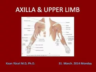
AXILLA & UPPER LIMB. Kaan Yücel M.D, Ph.D . 31. March . 2014 Monday. AXILLA ( ARMPIT). 6. Gateway to the upper limb An area of transition between the neck and the arm. BLOOD COMES ARTERIES BLOOD GOES VEINS MUSCLES,VESSELS, SWEAT GLANDS INNERVATED

E N D
AXILLA & UPPER LIMB Kaan Yücel M.D, Ph.D. 31. March. 2014 Monday
AXILLA (ARMPIT) 6 • Gateway to the upper limb • An area of transition between the neck and the arm.
BLOOD COMES ARTERIES BLOOD GOES VEINS MUSCLES,VESSELS, SWEAT GLANDS INNERVATED NERVES
Contents of the axilla • Axillary artery and its branches • Axillary vein and its tributaries • Lymph vessels and lymph nodes • Brachial plexus These structures are embedded in fat.
Axillary artery • Supplies the walls of the axilla & related regions. • Before: Subclavian artery After: Brachial artery • From lateral border of 1st rib • to • Inferior border of teres major
BRACHIAL PLEXUS a somatic nerve plexus - upperlimb formed by intercommunications amongventral rami of lower 4 cervical nerves ( C 5 - C 8) & T 1 responsible for motor innervation to all of muscles of upper limb exception trapezius.
BRACHIAL PLEXUS • supplies all of the cutaneous innervation of the upper limb • exception • area of the axilla (armpit) (intercostobrachialnerve) • an area just above thepoint of shoulder (supraclavicular nerves) • dorsal scapular area (cutaneous branches of dorsal rami) communicates with the sympatheticnervoussystem..
beginsin the neck and extends into the axilla. • Almost • all branches • of the brachial plexus • arise in the axilla • (after the plexus • has crossed the 1st rib). "Randy Travis Drinks Cold Beer"RootsTrunksDivisionsCords Branches· Alternatively: "Read The Damn Cadaver Book!"· Alternatively: "Real Texans Drink Coors Beer".
The parts of the brachial plexus, from medial to lateral, are roots, trunks, divisions, and cords. All major nerves that innervate the upper limb originate from the brachial plexus, mostly from the cords.
Long T h o r a c İ c nerve Dorsal scapular nerve Suprascapular nerve The nerve to subclavius muscle C5 Lateral pectoral nerve Musculocutaneous nerve Lateral root of median nerve Superior trunk C6 Lateral cord C7 Middle trunk Sup. & Inf. Subscapular nerves Thoracodorsal nerve Axillary nerve Radial nerve Posterior cord C8 Medial cord Inferior trunk T1 Medial pectoral nerve Medial cutaneous nerve of arm Medial cutanoues nerve of forearm Ulnar nerve Median nerve Red: Anterior division Blue: Posterior division "Randy Travis Drinks Cold Beer"RootsTrunksDivisionsCords Branches· Alternatively: "Read The Damn Cadaver Book!"· Alternatively: "Real Texans Drink Coors Beer".
shoulder Region of upper limb attachment to the trunk Proximal segment of limb overlaps parts of the trunk (thorax and back) and lower lateral neck. includes Pectoral Scapular Deltoid regions of the upper limb lateral part (greater supraclavicular fossa) of lateral cervical region. Overlies half of the pectoral girdle.
The pectoral (shoulder) girdle formed by: Scapulae and clavicles completed anteriorly by the manubrium of the sternum (part of the axial skeleton).
SUPERFICIAL POSTERIOR AXIOAPPENDICULAR MUSCLES EXTRINSIC SHOULDER MUSCLES Trapezius & latissimus dorsi DEEP POSTERIOR AXIOAPPENDICULAR MUSCLES EXTRINSIC SHOULDER MUSCLES Levatorscapulae& rhomboids
SCAPULOHUMERAL (INSTRINSIC SHOULDER) MUSCLES 6 scapulohumeral muscles Deltoid, teres major, supraspinatus, infraspinatus, subscapularis, and teres minor pass from scapula to humerus Act on the glenohumeral joint. All the intrinsic muscles but the deltoid and the subscapularis are muscles of the posterior scapular region.
4 muscles pass between the scapula and proximal end of humerus: POSTERIOR SCAPULAR REGION Supraspinatus Infraspinatus Teres minor Teres major
+ part of longhead of the triceps brachii, passesbetween the scapula and the proximal end of the forearm.
Supraspinatus & Infraspinatus • Originate from 2 large fossae, 1 above and 1 below the spine, on the posterior surface of the scapula. • Supraspinatus initiates abduction of the arm. • Infraspinatuslaterally rotates the humerus.
Teres minor • A cord-like muscle • Laterallyrotates the humerus • Component of the rotator cuff.
ROTATOR CUFF MUSCLES • 4 intrinsic shoulder muscles • Supraspinatus • Infraspinatus • Teres minor • Subscapularis (SITS muscles) Form a musculotendinous rotator cuff around the glenohumeral joint.
ROTATOR CUFF MUSCLES Functional exception: All except supraspinatus are rotators of the humerus Supraspinatus, besides being part of the rotator cuff, initiates and assists the deltoid in the first 15° of abduction of the arm.
Nerves The two major nerves of the posterior scapular region: Suprascapular & Axillary nerves originate from the brachial plexus in the axilla.
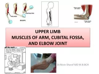
medial & lateral intermuscular septa Anteriorcompartment – flextheelbowjoint Posteriorcompartment- extendtheelbowjoint ARM Flexion Extension Pronation Supination
Anterior compartment of the arm coracobrachialis, brachialis, and biceps brachii muscles innervated predominantly by musculocutaneous nerve. Posterior compartment triceps brachii muscle innervated by radial nerve.
Brachial artery • Deep artery of the arm • (L. arteria profunda brachii) • Largest branch & most superior origin • Accompanies radial nerve along the radial groove • Terminates by dividing into middle & radial collateralarteries
2 main superficial veins of the arm cephalicand basilic veins. Cephalicvein – lateralside intoaxillaryvein Basilicvein- medialside Basilicvein+ Brachialveins Axillaryvein
4 main nerves pass through the arm: • Median • Ulnar • Musculocutaneous • Radial
1. Whichstructurespassbetweenthearm & forearm? Most major structures(nerves,veins,arteries) viacubital fossa,anterior to elbow joint Exception ulnar nerve posterior to the medial epicondyle of humerus
2. How is forearmdivided? pronation supination
3. Movements of theforearmmuscles? Muscles in theanteriorcompartment Flexthewrist & digits Pronatethehand Muscles in theposteriorcompartment Extendthewrist & digits Supinatethehand
4. Innervation of theforearmmuscles? Muscles of the anterior compartment Mainly by median nerve The one and a half exceptions by ulnar nerve Muscles of the posterior compartment All by radial nerve (directly or by its deep branch) Watch out, Median nerve @median plane of the forearm Ulnar nerve @ medial side
1. Layers of anteriorcompartmentforearmmuscles? SuperficialIntermediateDeep 3 muscles 4 muscles
2. Muscles of thesuperficiallayer? flexorcarpiradialis flexorcarpiulnaris palmarislongus pronatorteres twoheads Ulnarhead Humeralhead Medialepicondyle
2. Muscles of thesuperficiallayer? flexorcarpiradialis Medialepicondyle of humerus Base of metacarpals II & III 2. flexorcarpiulnaris Humeralhead: Medialepicondyle of humerus Ulnarhead: Olecranon & Posterior border of ulna •Pisiform& hamate •5th metacarpal 3. palmarislongus Medialepicondyle of humerus Flexor retinaculum &palmar aponeurosis 4. pronatorteres Humeralhead: Medialepicondyle& adjacentsupraepicondylarridge Ulnarhead: Coronoidprocess Lateral surface of radius
3. Muscles of theintermediatelayer? 3. ..the muscles of the intermediate and deep layers? flexor digitorum superficialis Humeroulnarhead • Medial epicondyle of humerus • Adjacent margin of coronoid process Radialhead Superior half of anterior border Shafts of middle phalanges of medial four digits
4. Muscles of thedeeplayer? 3. ..the muscles of the intermediate and deep layers? flexor pollicis longus flexor digitorum profundus pronator quadratus • Proximal¾ of medial& anteriorsurfaces of ulna • Interosseousmembrane • Anteriorsurface of radius • Adjacentinterosseousmembrane Distal ¼ of anterior surface of ulna Distal ¼ of anterior surface of radius Bases of distal phalanges of 4th &5th digits Bases of distal phalanges of 2nd &3rd digits Base of distal phalanx of thumb
5. Fxns of theanteriorcompartmentmuscles Flexion of forearm @ theelbowjoint Pronator teres Flexionof hand @ thewristjoint Flexorcarpiradialis et ulnaris- Palmarislongus Abduction (radialdeviation) of hand @ thewristjoint Flexorcarpiradialis Adduction (ulnardeviation) of hand @ thewristjoint Flexorcarpiulnaris Pronationof forearm Pronator teres – Pronatorquadratus
5. Fxns of theanteriorcompartmentmuscles Flexordigitorumsuperficialis • Flexesproximalinterphalangealjointsof theindex, middle, ring, & littlefingers • Flexesmetacarpophalangealjointsof thesamefingersandthewristjoint • Flexordigitorumprofundus • Flexesdistalphalanges4 & 5 at distalinterphalangealjoints • Flexes distal phalanges 2 and 3 at distal interphalangeal joints Flexorpollicislongus Flexesphalanges of thumb
6. Innervation of theanteriorcompartmentmuscles Allthemusclesbymediannerve Except1.5 musclesbyulnarnerve Flexorcarpiulnarisfull Flexordigitorumprofundusmedialhalf partassociated w/ring & littlefingers
7. Arteries in theanteriorcompartment of theforearm Brachialartery
8. Veins in theanteriorcompartment of theforearm deepvenouspalmararch in thehand
9. Mediannerve principalnerve no branches in the arm other than small twigs to the brachial artery. Its major branch in the forearm anterior interosseous nerve Leaves cubitalfossa by passing between 2 heads of pronator teres & humero-ulnar &radial heads of flexor digitorumsuperficialis
10. Ulnarnerve Enterstheanteriorcompartmentbypassingposteriorly around medialepicondyle of humerus & betweenhumeral& ulnarheads of flexorcarpiulnarismuscle Two small cutaneous branches palmar branch & dorsal branch
11. Radialnerve motor andsensory functionsin botharm & forearm but onlysensoryfunctions in thehand Superficial(sensory) deep to brachioradialis Deep(motor) between two heads of supinator
12. Lateral & medialcutaneousnerves of forearm Lateralcutaneousnerve of forearm Continuationof musculocutaneousnerve Medialcutaneousnerve of forearm Branch of medialcord of brachialplexus Posteriorcutaneousnerve of forearm Branch of radialnerve
1. What is the cubital fossa? An important area of transition between the arm and the forearm. seen superficially as a depression on the anterior aspect of the elbow. Deeply, it is a space filled with a variable amount of fat anterior to the most distal part of the humerus and the elbow joint.
2. What are the boundaries of the cubital fossa? Superiorly imaginary line connecting medial &lateral epicondyles. Mediallypronator teres. Laterallybrachioradialis.
3. Whatarethe contents of the cubital fossa? 1) Terminal part of the brachial artery,radial and ulnar arteries 2) Biceps brachii tendon 3) Median nerve 4) Radial nerve 5) (Deep) accompanying veins of the arteries
1. Which muscles are in the superficial layer? Lateral epicondyle of humerus Lateral epicondyle of humerus Brachioradialis Extensor carpi radialis longus Proximal part of supraepicondylar ridge of humerus Distal part of supraepicondylar ridge of humerus Extensor carpi radialis brevis Lateral epicondyle of humerus common extensor origin Lateral epicondyle of humerus common extensor origin Lateral epicondyle of humerus Extensor carpi ulnaris Extensor expansion (hood) of the 5th digit Extensor digitorum Olecranon and proximal posterior surface of ulna Extensor digiti minimi dorsal aspects of the bases of the middle and distal phalanges of the medial four digits Anconeus Lateral surface of distal end of radius proximal to styloid process Common originlateralepicondyle of the humerus Except for the brachioradialis and anconeus, extend as tendons into the hand. Base of 3rd metacarpal Base of 5th metacarpal Base of 2nd metacarpal Base of 2nd metacarpal Base of the 5th metacarpal Base of 3rdmetacarpals
Posterior surface of distal 1/3 of ulna and interosseous membrane Posterior surface of proximal halves of ulna, radius, and interosseous membrane 2. Which muscles are in the deep layer? Superficial (humeroulnar) head •lateral epicondyle of humerus •radial collateral and anular ligaments Deep (ulnar) head Supinator crest of the ulna Supinator Abductor pollicis longus Extensor pollicis longus Extensor pollicis brevis Base of 1st metacarpal Extensor indicis Posterior surface of middle third of ulna and interosseous membrane Dorsal surface of base of distal phalanx of thumb Lateral surface of radius superior to the anterior oblique line Extensor expansion (hood) of 2nd digit Posterior surface of distal third of radius and interosseous membrane Dorsal surface of base of proximal phalanx of the thumb Except for the supinator muscle, all these deep layer muscles originate from the posterior surfaces of the radius, ulna, and interosseous membrane and pass into the thumb and fingers.