Download
1 / 1
10 likes | 115 Views
How Does Baseline Airway Tone Modulate Bronchodilation During A Deep Inhalation?. D.A. AFFONCE 1 , A. GARRISON 2 , L.D. BLACK 1 , J.J. FREDBERG 3 , R. BROWN 4 , E. GARSHICK 2 AND K. LUTCHEN 1

E N D
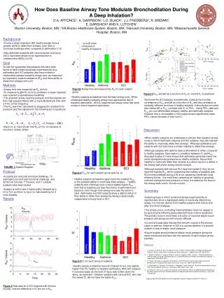
How Does Baseline Airway Tone Modulate Bronchodilation During A Deep Inhalation? D.A. AFFONCE1, A. GARRISON2, L.D. BLACK1, J.J. FREDBERG3, R. BROWN4, E. GARSHICK2 AND K. LUTCHEN1 1Boston University, Boston, MA; 2VA Boston Healthcare System, Boston, MA; 3Harvard University, Boston, MA; 4Massachusetts General Hospital, Boston, MA Background • During a deep inspiration (DI) healthy people have a greater ability to dilate their airways, even after a bronchial challenge when compared to asthmatics (1,2) • Non asthmatic subjects with cervical spinal cord injury (SCI) have been shown to be hyperreactive to methacholine (MCh) (3,4,5) * p<0.05 when compared to healthy at baseline * * * * * * Goal To test the hypothesis that subjects that have been found to have enhanced airway hyperreactivity (e.g. asthmatics and SCI subjects) also have a distinct relationship between baseline airway tone, as measured by respiratory system resistance (Rrs) and there ability to dilate their airways with a DI Post Challenge Post Challenge Thoracic SCI Cervical SCI Lumbar SCI Baseline Baseline All SCI Methods Figure3 Airway tone as measured by Rrs for each subject group • Airway tone was measured as Rrs at 8 Hz • To measure Rrs@8 Hz an 8 Hz oscillation is super imposed over a person’s spontaneous breathing • The acquired airway opening pressure and flow signals are then high passed filtered with a 4 pole Butterworth filter with a 4 Hz corner frequency • The Rrs was then calculated by plugging the isolated 8 Hz pressure and flow signals into the following set of equations: Figure 6 Rmin and ΔR as a function of Rrs,IC, and FEV1 % predicted • Healthy subjects at baseline have the least airway tone. When challenged healthy subjects airway tone approached that of baseline asthmatics. All SCI subjects had airway tones that were similar to that of baseline asthmatics. • Note how the SCI subjects and asthmatic subjects have similar correlations of Rmin and ΔR as a function of Rrs and this correlation is markedly different from that of healthy subjects. It should also be noted that neither ΔR or Rmin correlate with IC or FEV1. Using FEV1 data and Rmin you can differentiate between healthy and asthmatic subjects. However this is not possible in SCI subjects have significantly lower FEV1 values because of their low IC. * • Black et. al. have shown that Rrs at TLC is indicative of maximum airway caliber * Discussion * * * * • When healthy subjects are challenged to elevate their baseline airway tones to that of Asthmatic subjects and SCI subjects, they still maintain the ability to maximally dilate their airways. Whereas asthmatics and subjects with SCI both have a similar inability to dilate their airways • Although subjects with asthma have a diminished IC when compared to healthy subjects there was no correlation between IC and Rmin. Also past studies (1) have shown that asthmatics generate nearly the same transpulmonary pressures as healthy subjects. Hence their inability to maximally dilate their airways is a direct result of a defect in the airway wall and/or airway smooth muscle • Although subjects with cervical SCI have the lowest IC they do not have the highest Rmin which means that the inability of subjects with SCI to bronchodilate during a DI is not caused by insufficient local tethering forces. It is most likely caused by an inability to generate sufficient transpulmonary pressure or there is a defect at the level of the airway walls and/or smooth muscle * Post Challenge Post Challenge Thoracic SCI Lumbar SCI Cervical SCI Baseline Baseline All SCI FIGURE 1:System Diagram Protocol Figure 4 Rmin for each subject group (see fig. 2) • 6 healthy pre and post bronchial challenge, 10 asthmatics pre and post bronchial challenge, and 22 SCI (8 Cervical, 7 Thoracic, and 7 Lumbar) subjects have been tested • Subject is told to take 5 tidal breaths followed by a DI to TLC and then to return to tidal breathing for 5 more breaths • Healthy subjects at baseline again have the smallest Rmin, or the greatest ability to maximally dilate airways . Healthy subjects post challenge have a value slightly higher Rmin than that at baseline, but less than those of asthmatics and baseline, even though their airway tone was the same to start. Asthmatics and SCI subjects show a distinct defect in their ability to dilate their airways by taking a deep breath, independent of injury level in SCI. Summary • Subject groups in which enhanced airway hyperreactivity is reported also show a depressed ability to maximally dilate there airway in a manner distinct from healthy subjects both before and after bronchial challenge • The primary force controlling maximal dilation does not appear to be parenchyma tethering associated with local volume expansion. The primary force is more likely a function of maximal elastic recoil pressure and airway smooth muscle stiffness • Current and past data indicate that smooth muscle is the primary culprit in asthma. However in SCI it is unclear whether it is smooth muscle or loss of elastic recoil pressure • Future studies should measure elastic recoil pressure during the same maneuvers and also test the reactivity of each subject group explicitly Results * * * * Post Challenge Post Challenge Thoracic SCI Lumbar SCI Cervical SCI Baseline References Baseline All SCI • A. Jensen, H. Atileh, B. Suki, E. Ingenito, and K. Lutchen. Airway Caliber in Healthy and Asthmatic Subjects: Effects of Bronchial Challenge and Deep Inspiration. J Appl Physiol 2001 96:506-515 • L. Black, R. Dellaca, K. Jung, H. Atileh, E. Israel, E. Ingenito, and K. Lutchen. Tracking Variations in Airway Caliber by Using Total Respiratory Vs. Airway Resistance in Healthy and Asthmatic Subjects. J. Appl Physiol 2003 95:511-518 • E. Singas, M. Lesser, A. Spungen, W. Bauman, and P Almenoff. Airway Hyperresponsiveness to Methacholine in Subjects With Spinal Cord Injury. Chest Oct. 1996 110(4):911-915 • P. Dicpinigaitis, A. Spungen, W. Bauman, A. Absgarten, and P. Almenoff. Bronchial Hyperresponsiveness After Cervical Spinal Cord Injury. Chest April 1994 109(4):1073-1076 • D. Grimm, R. DeLuca, M. Lesser, W. Bauman, and P. Almenoff. Effects of GABA-B Agonist Baclofen on Bronchial Hyperreactivity to Inhaled Histamine in Subjects with Dervical Spinal Cord Injury. Lung 1997 175:333-341 Figure 5 IC for each group of subjects Rmin • Healthy people at baseline have the highest IC but only slightly higher then PC healthy or baseline asthmatics. With SCI subjects IC became larger as the level of injury was further down the spine, as expected. However subjects with cervical SCI, who had the lowest IC, did not have the higher Rmin Supported by NIH HLB 62269, the DVA Cooperative Studies Program and NIH R01 HD42141 Figure 2 Raw data for 2 SCI subjects with thoracic injuries, note the difference in IC but not in Rmin