Download
1 / 19
210 likes | 440 Views
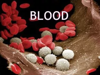
Blood. Blood. Consists of blood cells & plasma Blood cells = Erythrocytes (RBC’s), Leukocytes (WBC’s), & Thrombocytes (Platelets) Blood is 55% plasma & 45% blood cells Woman has ≈ 5 liters Man has ≈ 6 liters. Functions. Transport Nutrients, waste, hormones, enzymes, O 2 & CO 2

E N D
Blood • Consists of blood cells & plasma • Blood cells = Erythrocytes (RBC’s), Leukocytes (WBC’s), & Thrombocytes (Platelets) • Blood is 55% plasma & 45% blood cells • Woman has ≈ 5 liters • Man has ≈ 6 liters
Functions • Transport • Nutrients, waste, hormones, enzymes, O2 & CO2 • Regulation of body temp due to high volume of H2O in plasma • Helps regulate body pH • Helps regulate water content of cells/Osmosis • Clotting: prevents fluid loss • Protection against pathogens (immune response, production of antibodies, destruction of bacteria/viruses, removal of cellular debris & allergic reactions)
Plasma Composition • 91% water • 7% proteins: • Albumin: maintains osmotic pressure & water balance • Globulins: antibodies, complements (immune response) & transport molecules • Fibrinogens: important role in clotting • 2% solutes: • Ions, nutrients, waste products, gases, enzymes & hormones
Erythrocytes (RBC’s) • 95% of blood cell volume; biconcave disks • No nucleus, simple structures, don’t divide & live ≈ 120 days • Composed of a network of protein called stroma, cytoplasm, lipids (cholesterol) & hemoglobin (red pigment ≈ 33% of cell’s volume)
Erythrocytes (RBC’s) • Function: • Transport O2 & CO2→→ Hemoglobin allows this • Hemoglobin: • Globin = protein • Heme = pigment containing 4 iron atoms • Iron combines with O2 in the lungs & releases it in tissues; Bright red in color • Globin at tissues combines with CO2 & releases it at lungs; Dark red in color
Leukocytes (WBC’s) • 2 subcategories: Granular & Nongranular • Have nuclei & no pigment; larger than RBC’s • General function in immune response: • Combat inflammation & infection • Can leave the blood stream & move into tissues via ameboid movement • Phagocytosis: “cell eating”
Granular leukocytes: have granules in cytoplasm • Neutrophils: 60% - 70% of WBC’s • Most active in WBC’s response to tissue destruction by bacteria • Stay in blood for 12 hours & then move to tissues where they phagocytize (eat) foreign substances • Secrete enzyme Lysozyme that destroys certain bacteria • Pus contains dead neutrophils, cell debris & fluids
Granular leukocytes • Eosinophils: 2% - 4% of WBC’s • Combat irritants (pollen, dust, pet dander, etc) that causes allergies • Produce antihistamines • Basophils: 0.5% - 1% of WBC’s • Involved in allergic reactions • Releases heparin (anticoagulant), histamine (inflammatory substance) & serotonin (a vasoconstrictor)
Nongranular leukocytes: no granules in cytoplasm • Monocytes: 3% - 8% of WBC’s • Phagocytotic: eat bacteria, dead cells &/or cellular debris • Largest; after they leave blood & enter tissue, they increase in size & are called Macrophages • Lymphocytes: 20% - 25% of WBC’s • Production of antibodies & play important role in immune response • Smallest; several types: B & T lymphocytes • Control cancer cells, destroy microorganisms & reject foreign tissues
Thrombocytes (platelets) • Disk-shaped cellular fragments with a nucleus • Prevent fluid loss when blood vessels are damaged
Clotting Mechanism • When larger blood vessels are damaged, clotting mechanism takes over • Cut vessel is rough & irregular shaped • 3 stages to clotting/coagulation • Rough surface of vessel causes platelets to clump together at the site of the injury • Tissue releases thromboplastin which produces prothrombin activator • Requires Ca2+, certain proteins & phospholipids
Clotting Mechanism • Ca2+ & prothrombin activator converts prothrombin into thrombin • Soluble fibrinogen is converted into insoluble fibrin • Thrombin catalyzes the reaction • Fibrin forms long threads that act like a net = CLOT • Clot forms & traps blood cells & platelets in the fibrin threads & bleeding stops
Clotting Mechanism • Syneresis: clot retraction; tightening of clot so wound gets smaller & smaller • Serum (blood plasma minus clotting factors) surrounds wound under clot & hemorrhage is stopped • Blood vessel repairs itself • Fibronolysis occurs: blood clot dissolves
Problems in clotting • Build up of cholesterol mass (Plaque) on smooth walls of UNDAMAGED blood vessels can cause clot formation • Called Thrombosis & clot is called a Thrombus • Thrombus may dissolve or a piece can dislodge & get transported in blood = Embolus • Embolus can get stuck in a vessel & cut-off circulation = Embolism • If tissues are killed = Infarction
Blood groups • Agglutination: clumping of RBC’s, A.K.A transfusion reaction • Caused by reaction between antibodies in plasma & surface antigens on RBC’s • Caused by mismatched blood types • Headache, difficulty breathing, face flushed, pain in neck, chest & lower back, jaundice & kidney failure
Abo blood groups • Presence or absence of antigens on RBC surface: antigen A & antigen B • Inherited; 4 possible antigen combinations: • A only: Type A • B only: Type B • A & B : Type AB • Neither A nor B: Type O • Antibodies are formed during infancy against the ABO antigens NOT present on our own RBC’s • Type A: antibody anti-B • Type B: antibody anti-A • Type AB: neither antibody → • Type O: both anti-A & anti-B Universal Recipient Universal Donor
Rh blood groups • Inherited; named after Rhesus monkey where antigen 1st discovered • If antigen D is found on RBC, the blood is Rh positive • If the RBC lacks the antigen, blood is Rh negative • Anti-Rh antibodies only develop after initial exposure to Rh-positive blood • Rh-negative person receives transfusion from Rh-positive person = no reaction (1st time) but anti-Rh antibodies form • If 2nd exposure happens, agglutination occurs


