Download

1 / 58
620 likes | 1.24k Views
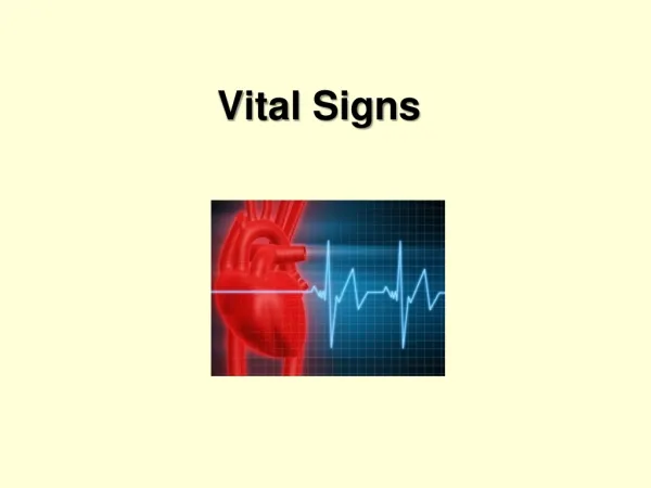
Introduction to Vital Signs and Basic Laboratory Tests. Joel N. Kniep, M.D. Dept. of Pathology. Objectives. Introduce vital signs and their use in clinical practice Introduce basic laboratory tests and their use in clinical practice Discuss normal values and test interpretation.

E N D
Introduction to Vital Signs and Basic Laboratory Tests Joel N. Kniep, M.D. Dept. of Pathology
Objectives • Introduce vital signs and their use in clinical practice • Introduce basic laboratory tests and their use in clinical practice • Discuss normal values and test interpretation
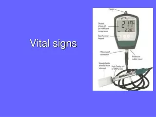
Clinical Vital Signs (Vitals) • Temperature • Pulse rate • Respiration rate (RR) • Blood pressure (bp)
Temp • Measure of body’s core temp (temp of internal organs) • in ° F (or ° C) • Locations: oral, rectum, axilla, ear • Rectal = 0.5 – 0.7° F higher than oral temp • Axilla = 0.3 – 0.4° F lower than oral temp • Normal: 97.8 – 99° F (36.5 – 37.2° C) • Critical: > 98.6° F orally or 99.8° F rectally (pyrexia [fever]); < 95° F (hypothermia)
Pulse rate • Heart rate (HR) or number of heart beats/min • Normal: 60 – 100/min • ↑ (tachycardia): ↑ Na+ intake, ↓ Na+ loss, Excessive free body H2O loss • ↓ (bradycardia): ↓ Na+ intake, ↑ Na+ loss, ↑ free body H2O
RR • Number of breaths/min • At rest • Also note breathing effort or difficulty • Normal: 15 – 20/min • Critical: < 12 or > 25 • ↑ (hyperventilation): ↑ Na+ intake, ↓ Na+ loss, Excessive free body H2O loss • ↓ (hypoventilation): ↓ Na+ intake, ↑ Na+ loss, ↑ free body H2O
Bp • Measures the force of blood against the arterial vessel walls • Measured while seated, after resting for 5 mins, arm resting @ heart level (if possible) • Reported as a fraction (systolic/diastolic) & consists of 2 separate measurements: • Systolic – pressure within artery during cardiac contraction • Diastolic – pressure within artery during cardiac relaxation and filling • Normal: < 120 mm Hg systolic and < 80 mm Hg diastolic • Critical: > 220 mm Hg systolic or > 125 mm Hg diastolic • ↑ (hypertension [htn]): ↑ Na+ intake, ↓ Na+ loss, Excessive free body H2O loss • ↓ (hypotention): ↓ Na+ intake, ↑ Na+ loss, ↑ free body H2O
Complete Blood Count (CBC) • Provides information on cellular components of blood • Includes RBC count, Hemoglobin (Hgb), Hematocrit (Hct), RBC indices, White blood cell (WBC) count and differential, Platelet count
Total WBCs (leukocytes) • Measurement of total WBC count • Consists of total # of WBCs/mm3 of peripheral venous blood • Part of “routine” testing • Useful for evaluation of infection, neoplasm, allergy & immunosuppression • Normal: 4,000 – 10,000/mm3 • Critical: < 2,500 or > 30,000/mm3 • ↑ (leukocytosis): infection, malignancy, trauma, stress, hemorrhage, tissue necrosis, inflammation, dehydration, thyroid storm • ↓ (leukopenia): drug toxicity, bone marrow failure, overwhelming infections, dietary deficiency, congenital marrow aplasia, bone marrow infiltration, autoimmune disease, hypersplenism
Erythrocyte count (RBC) • Measures # of circulating RBCs/mm3 of peripheral venous blood • Direct measure of RBC count • Part of “routine” testing and anemia evaluation • Normal: 3.5 – 5.5 x 10⁶/μL • ↑: erythrocytosis, congenital heart disease, severe COPD, polycythemia vera, severe dehydration, hemoglobinopathies • ↓: anemia, hemoglobinopathy, hemorrhage, bone marrow failure, renal disease, leukemia, prosthetic valves, normal pregnancy, multiple myeloma, Hodgkin disease, lymphoma, dietary deficiency
Hgb • Measures total amount of Hgb in blood • Indirect measure of RBC count • Part of “routine” testing and anemia evaluation • Normal: 12 – 15 g/dL • Critical: < 5 or > 20 g/dL • ↑: erythrocytosis, congenital heart disease, severe COPD, polycythemia vera, severe dehydration ↓: anemia, hemoglobinopathy, hemorrhage, bone marrow failure, renal disease, leukemia, prosthetic valves, normal pregnancy, multiple myeloma, Hodgkin disease, lymphoma, dietary deficiency
Hct • Measure of RBC percent of total blood vol • Indirect measure of RBC # & volume • Part of “routine” testing and anemia evaluation • Normal: 36 – 48% • Critical: < 15% or > 60% • ↑: erythrocytosis, congenital heart disease, severe COPD, polycythemia vera, severe dehydration • ↓: anemia, hemoglobinopathy, hemorrhage, bone marrow failure, renal disease, leukemia, prosthetic valves, normal pregnancy, multiple myeloma, Hodgkin disease, lymphoma, dietary deficiency
RBC indices • Measures size and hgb content of RBCs • Used to classify anemias • Includes Mean corpuscular volume (MCV), mean corpuscular hemoglobin (MCH), mean corpuscular hemoglobin concentration (MCHC), red blood cell distribution width (RDW)
MCV • Measure of average volume/size of single RBC • MCV = Hct (%) x 10/RBC (million/mm3) • Useful in anemia classification • Normal: 80 – 100 mm3 • ↑ (macrocytic): pernicious anemia (vit B12 deficiency), folic acid deficiency, antimetabolic therapy, alcoholism, chronic liver disease, hypothyroidism • Normocytic: bone marrow failure/replacement, acute blood loss, chronic diseases, hemolytic anemias • ↓ (microcytic): Fe deficiency anemia, thalassemia, anemia of chronic illness
MCH • Measure of average amount of hgb within a single RBC • MCH = Hgb (g/dL) x 10/RBC (million/mm3) • Provides little additional info to other indices • Normal: 24 – 32 pg • ↑: macrocytic anemias • ↓: microcytic anemia, hypochromic anemia
MCHC • Measure of average [hgb] within a single RBC • MCHC = Hgb (g/dL) x 100/Hct (%) • 37 g/dL = maximum Hgb able to fit into an RBC (cannot be hyperchromic) • Normal (normochromic): 32 – 36 g/dL • ↑: spherocytosis, intravascular hemolysis, cold agglutinins • ↓ (hypochromic): Fe deficiency anemia, thalassemia
RDW • Measure of variation of RBC size (indicator of degree of anisocytosis) • Useful in anemia classification • Normal: variation of 11.5 – 16.9% • ↑: Fe deficiency anemia, vit B12 or folate deficiency anemia, hemoglobinopathies, hemolytic anemias, posthemorrhagic anemias
Platelet count • Measurement of platelets (thrombocytes) • Consists of actual # of platelets/mm3 of peripheral venous blood • Part of “routine” testing • Useful for evaluation of petechiae, spontaneous bleeding, increasingly heavy menses or thrombocytopenia • Useful for monitoring discourse/therapy of thrombocytopenia/bone marrow failure • Normal: 150,000 – 400,000/mm3 • Critical: < 50,000 or > 1,000,000/mm3 • ↑ (thrombocytosis): malignant disorders, polycythemia vera, postsplenectomy syndrome, rheumatoid arthritis, Fe deficiency anemia • ↓ (thrombocytopenia): Hypersplenism, hemorrhage, immune thrombocytopenia, leukemia & other myelofibrosis disorders, TTP, DIC, SLE, chemotherapy, pernicious anemia
WBC definitions • Leukocytosis – abnormally large number of leukocytes; generally indicated by WBC count of ≥ 10,000 cells/mm3 • Lymphocytosis – form of actual or relative leukocytosis due to increase in numbers of lymphocytes • Left shift – increase in the number of immature neutrophils (bands/stabs) found in the blood
WBC differential • Measurement of percentage of each WBC type in specimen • Useful for infection, neoplasm, allergy & immunosuppression evaluations • Normal: Neutrophils (50 – 70%), Lymphocytes (20 – 40%), Monocytes (2 – 8%), Eosinophils (0 – 5%), Basophils (0 – 2%) • ↑: refer to individual cell types on chart • ↓: refer to individual cell types on chart
Basic Metabolic Panel (BMP) • Measures electrolytes, chemicals, metabolic end products & substrates • Consists of Glucose, Blood Urea Nitrogen (BUN), Creatinine, Na+, K+, Cl-, Bicarbonate (HCO3-), Ca2+
Glucose • Direct measure of blood glucose • Commonly used to evaluate diabetic pts • Part of “routine” testing • Normal: 70 - 100 mg/dL • Critical: < 50 and > 400 mg/dL (♂) or < 40 and > 400 mg/dL (♀) • ↑ (hyperglycemia): DM, acute stress response, Cushing syndrome, pheochromocytoma, chronic renal failure, acute pancreatitis, acromegaly, corticosteroid therapy • ↓ (hypoglycemia): insulinoma, hypothyroidism, hypopituitarism, Addison disease, extensive liver disease, insulin overdose, starvation
BUN • Measures urea nitrogen in blood • End product of protein metabolism (produced in liver) • Indirect measure of renal function & glomerular function (excretion) • Measure of liver metabolic function • Part of routine labs • Usually interpreted along with Cr (less accurate than Cr for renal disease) • Normal: 6 -21 mg/dL • Critical: > 100 mg/dL • ↑: prerenal causes, renal causes, postrenal azotemia • ↓: liver failure, overhydration because of SIADH, neg nitrogen balance, pregnancy, nephrotic syndrome
Creatinine • Measures serum creatinine • Catabolic product of creatine phosphate (skeletal muscle contraction) • Excreted entirely by kidneys → direct measure of renal function • Minimally affected by liver function • Elevation occurs slower than BUN • Doubling ≈ 50% reduction in GFR • Normal: 0.44 – 1.03 mg/dL • Critical: > 4 mg/dL • ↑: diseases affecting renal function (glomerulonephritis, pyelonephritis, ATN, urinary tract obstruction, reduced renal blood flow, diabetic nephropathy, nephritis), rhabdomyolysis, acromegaly, gigantism • ↓: debilitation, decreased muscle mass
Na+ • Measures serum sodium level • Major cation in EC space • Balance between dietary intake and renal excretion • Normal: 136 – 146 mEq/L • Critical: < 120 or > 160 mEq/L • ↑ (hypernatremia): ↑ Na+ intake, ↓ Na+ loss, Excessive free body H2O loss • ↓ (hyponatremia): ↓ Na+ intake, ↑ Na+ loss, ↑ free body H2O
K+ • Measures serum potassium level • Major cation within cell • Normal: 3.4 – 5.2 mEq/L • Critical: < 2.5 or > 6.5 mEq/L • ↑ (hyperkalemia): excessive intake, acidosis, acute/chronic renal failure, Addison disease, hypoaldosteronism, infection, dehydration • ↓ (hypokalemia): deficient intake, burns, hyperaldosteronism, Cushing syndrome, RTA, licorice ingestion, alkalosis, renal artery stenosis
Cl- • Measures serum chloride level • Major anion in EC space • Helps maintain electrical neutrality; follows sodium • Normal: 98 – 108 mEq/L • Critical: < 80 or > 115 mEq/L • ↑ (hyperchloremia): dehydration, metabolic acidosis, RTA, Cushing syndrome, renal dysfunction, respiratory alkalosis, hyperparathyroidism • ↓ (hypochloremia): overhydration, SIADH, CHF, chronic respiratory acidosis, metabolic alkalosis, Addison disease, Aldosteronism, vomiting/prolonged gastric suction, hypokalemia
HCO3- • Measures CO2 content of blood • Major role in acid-base balance • Regulated by kidneys • Used to evaluate pt pH status & electrolytes • Normal: 22 – 32 mEq/L • Critical: < 6 mEq/L • ↑: severe vomiting, high-volume gastric suction, aldosteronism, mercurial diuretic use, COPD, metabolic alkalosis • ↓: chronic diarrhea, chronic loop diuretic use, renal failure, DKA, starvation, metabolic acidosis, shock
Ca2+ • Measures serum calcium level • Direct measurement • Used to evaluate parathyroid function & Ca metabolism • Used to monitor renal failure, renal transplantation, hyperparathyroidism, various malignancies, & Ca level when giving large-volume blood transfusions • Normal: Total = 8.3 – 10.3 mg/dL, Ionized = 4.5 – 5.6 mg/dL • Critical: Total < 6 or > 13 mg/dL, Ionized < 2.2 or > 7 mg/dL • ↑ (hypercalcemia): hyperparathyroidism, bone mets, Paget disease of bone, prolonged immobilization, milk-alkali syndrome, vit D intoxication, hyperthyroidism • ↓ (hypocalcemia): hypoparathyroidism, renal failure, rickets, vit D deficiency, osteomalacia, pancreatitis, alkalosis, malabsorption, fat embolism
Comprehensive Metabolic Panel (CMP) • Includes all components of BMP plus Albumin, Total protein, Alkaline phosphatase (ALP), Alanine aminotransferase (ALT), Aspartate aminotransferase (AST) and Bilirubin
Albumin • Measures amount of albumin in blood • Formed within liver & comprises 60% of total protein in blood • Maintains colloidal osmotic pressure & transports blood constituents • Measure of both hepatic function and nutritional state • Normal: 3.5 – 5 g/dL • ↑: dehydration • ↓: malnutrition, pregnancy, liver disease, protein-losing enteropathies, protein-losing nephropathies, 3rd space losses, overhydration, ↑ capillary permeability, inflammatory disease, familial idiopathic dysproteinemia
Total Protein • Measures total protein in blood • Combination of prealbumin, albumin & globulins • Normal: 6.4 – 8.3 g/dL
ALP • Measures serum ALP concentration • Detect & monitor liver and bone disease • Normal: 30 -120 units/L • ↑: 1° cirrhosis, intrahepatic/extrahepatic biliary obstruction, 1°/metastic liver tumor, hyperparathyroidism, Paget disease, normal growing bones in children, bone mets, RA, MI, sarcoidosis, healing fracture, normal pregnancy, intestinal ischemia or infarction • ↓: hypophosphatemia, malnutrition, milk-alkali syndrome, pernicious anemia, scurvy
ALT • Found predominantly in liver • Injury/disease to parenchyma → release into blood • ID & monitor hepatocellular diseases of liver • If jaundiced, implicates liver rather than RBC hemolysis • Normal: 4 – 36 international units/L @ 37°C • Sig ↑: hepatitis, hepatic necrosis, hepatic ischemia • Mod ↑: cirrhosis, cholestasis, hepatic tumor, hepatotoxic drugs, obstructive jaundice, severe burns, trauma to striated muscle • Mild ↑: myositis, pancreatitis, MI, infectious mono, shock
AST • Found in highly metabolic tissue (cardiac & skeletal muscle, liver cells) • Disease/injury → lysing of cells & release into blood • Elevation proportional to # of cells injured • Used for evaluation of suspected coronary artery disease or hepatocellular disease • Normal: 0 – 35 units/L • ↑: heart diseases, liver diseases, skeletal muscle diseases • ↓: acute renal disease, beriberi, DKA, pregnancy, chronic renal dialysis
Bilirubin • Measures level of total bilirubin in blood • End product of RBC metabolism (RBCs → Hgb → Heme (+ globin) → Biliverdin → Bilirubin (unconjugated/indirect) → Bilirubin (conjugated/direct) • Component of bile • Consists of conjugated (direct) & unconjugated (indirect) bilirubin • Used to evaluate liver function; hemolytic anemia workup in adults & jaundice in newborns • Jaundice occurs when total bilirubin > 2.5 mg/dL • Normal: 0.3 – 1 mg/dL • Critical: > 12 mg/dL
Unconjugated bilirubin • Measures level of indirect bilirubin in blood • Normal: 0.2 – 0.8 mg/dL • ↑: erythroblastosis fetalis, transfusion rxn, sickle cell anemia, hemolytic jaundice, hemolytic anemia, pernicious anemia, large-volume blood transfusion, large hematoma resolution, hepatitis, cirrhosis, sepsis, neonatal hyperbilirubinemia, Crigler-Najjar syndrome, Gilbert syndrome
Conjugated bilirubin • Measures level of direct bilirubin in blood • Produced by conjugating glucuronide w/ unconjugated/indirect bilirubin in liver • Normal: 0.1 – 0.3 mg/dL • ↑: gallstones, extrahepatic duct obstruction, extensive liver mets, cholestasis from drugs, Dubin-Johnson syndrome, Rotor syndrome
Urinary Analysis (UA) • Provides information about kidneys & other metabolic processes • Used for diagnosis, screening & monitoring • Frequently used to test for urinary tract infections (UTIs)
UA Normal Values • Appearance: clear • Color: amber yellow • Odor: aromatic • pH: 4.6 – 8 • Protein: 0 – 8 mg/dL • Specific gravity: 1.005 – 1.030 • Leukocyte esterase: negative • Nitrites: none • Ketones: none
UA Normal Values cont. • Bilirubin: none • Urobilinogen: 0.01 – 1 Ehrlich unit/mL • Crystals: none • Casts: none • Glucose: negative • White Blood Cells: 0 – 4/low-power field • WBC casts: none • Red Blood Cells (RBCs): ≤ 2 • RBC casts: none
Urinary Protein • Used to monitor kidney function • Normally not present in normal kidney due to size barrier in glomerulous • Normally tested by dipstick method, quantification requires 24-hour urine collection • Presence (proteinuria) can indicate nephrotic syndrome, multiple myeloma or complications of DM, glomerulonephritis, amyloidosis
Urinary Glucose • Glucosuria – presence of glucose in urine • Reflection of serum glucose levels • Helpful in monitoring DM therapy • Renal glucose reabsorption threshold = 180 mg/dL (in proximal renal tubules) • Not always abnormal • Can occur after a high-carbohydrate meal or IV dextrose fluids • Can occur in diseases affecting renal tubules; genetic defects of metabolism & glucose excretion • ↑: DM & other causes of hyperglycemia, pregnancy, renal glycosuria, Fanconi syndrome, Hereditary defects in metabolism of other reducing substances, ↑ ICP, nephrotoxic chemicals
Urinary Leukocyte esterase • Screen to detect leukocytes in urine (dipstick method) • Presence indicates UTI • 90% accurate
Urinary Ketones • End products of fatty acid catabolism • Examples: β-hydroxybutyric acid, acetoacetic acid, acetone • Associated with poorly controlled diabetes • Used to evaluate ketoacidosis associated w/ alcoholism, fasting, starvation, high-protein diets, isopropanol ingestion
Urinary Nitrites • Screen for UTI (dipstick method) • Test based on chemical rxn by bacterial reductase (reduces nitrate to nitrite) • 50% accurate • Enhances leukocyte esterase sensitivity
Urinary Casts • Hyaline – conglomerations of protein; indicative of proteinuria; few = normal especially after exercise • Cellular – conglomerations of degenerated cells • Granular – glomerular disease • Fatty – nephrotic syndrome • Waxy – chronic renal disease • Epithelial cells & casts (renal tubular casts) • WBCs & casts – acute pyelonephritis • RBCs & casts – glomerular diseases
Cerebral Spinal Fluid (CSF) Analysis • Collected via lumbar puncture (LP) • Useful for the diagnosis of 1° or metastatic brain/spinal cord neoplasm, cerebral hemorrhage, meningitis, encephalitis, degenerative brain disease, autoimmune diseases w/ CNS involvement, neurosyphilis, demyelinating diseases
CSF analysis Normal Values • Opening pressure: <20 cm H2O • Color: clear & colorless • Blood: none • RBCs: 0 • WBCs: 0 – 5 cells/μL • Neutrophils: 0 – 6% • Lymphocytes: 40 – 80% • Monocytes: 15 – 45%
CSF analysis Normal Values cont. • Protein: 15 – 45 mg/dL • Glucose: 50 – 75 mg/dL or 60 – 70% of blood glucose level