Function of the heart
Function of the heart. Chapter 17. Cardiac Cycle. Sequence of events that occurs during one heartbeat Coordinated contraction and relaxation of the chambers of the heart Systole- contraction of myocardium Diastole- relaxation of myocardium. Systole & Diastole. Systole

Function of the heart
E N D
Presentation Transcript
Function of the heart Chapter 17
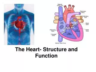
Cardiac Cycle • Sequence of events that occurs during one heartbeat • Coordinated contraction and relaxation of the chambers of the heart • Systole- contraction of myocardium • Diastole- relaxation of myocardium
Systole & Diastole • Systole • Contraction of heart muscle forces blood out of the chamber • Diastole • Relaxation of heart muscle allows the chamber to fill with blood • Atrial and ventricular activity are closely coordinated: atrial systole = ventricular diastole
Three Stages of Cardiac Cycle • Atrial Systole • Atria contract; pump blood into ventricles • AV valves open, ventricles relaxed • Ventricular Systole • Ventricles contract; pushes AV valves closed; pushes semilunar valves open • Blood pumped to pulmonary artery & aorta
Three Stages of Cardiac Cycle • Diastole • Brief time when both atria and ventricles are relaxed • Blood flows into atria; some blood flows passively into ventricles • Diastole is a “filling” period • Cycle repeats itself starting with atrial contraction again
Which of the following occurs during ventricular diastole? • The ventricles fill with blood. • The atrioventricular valves close. • The ventricles pump blood into the great vessels. • The semilunar valves open.
Cardiac Cycle • Cardiac cycle is repeated with every heartbeat; if heart rate is 70 bpm, then cardiac cycle lasts approx. 0.8 sec; diastole lasts approx. 0.4 sec • If heart rate increases, diastole shortens- can impact cardiac function. How? • Decreased filling time reduces the amount of blood that enters the ventricles; and coronary blood flow occurs during diastole
Autonomic Control of the Heart • If cardiac cells can initiate cardiac impulses, why are autonomic nerves needed? • Affect the rate at which cardiac impulses are fired • Affects how fast the impulses travel through the heart • Affects how forcefully the heart contracts
ANS • The autonomic nervous system allows the heart to respond to increased oxygen demand by increasing the rate and force of cardiac contraction.
Autonomic Wiring • Sympathetic • Supply the SA node, AV node and ventricular myocardium • Parasympathetic • Vagus nerve • SA node and AV node (does not innervate the ventricles)
Autonomic Firing • Sympathetic stimulation • Increases SA node activity ( HR) • Increases speed of impulse (from SA node to His-Purkinje) • Increases strength of contraction
Important points to remember • Excessive sympathetic activity leads to “fight or flight” response (panic causes racing and pounding heart) • May be involved in certain illnesses- shock, heart failure (need to treat with drugs that reduce excessive sympathetic firing)
Important points to remember • Causes tachydysrhythmias • Nurses often give drugs that mimic or block sympathetic activity • Drugs that mimic sympathetic activity increase HR and force of contraction (epinepherine & dopamine); called sympathomimetic drugs • Drugs that inhibit SNS effects are called sympatholytic drugs (clonidine)
Autonomic Firing • Paraympathetic stimulation • Decreases SA node activity ( HR) • Decreases the speed of cardiac impulses from SA to AV node • Does not affect strength of myocardial contraction (no innervation of ventricles)
Important points to remember • Parasympathetic effects are exerted by the vagus nerve • In the resting heart, the vagus nerves slows the firing of the SA node (SA node wants to fire at 90 bpm, vagus nerve keeps it around 70) • Excessive vagal discharge can be caused by different things, including certain drugs(digoxin) and conditions (MI)
Important points to remember • Excessive vagal discharge causes bradycardia (<60 bpm); it also increases the likelyhood of lethal dysrhythmias • Vagal stimulation can also slow conduction through the heart, leading to potentially lethal heart blocks
Important points to remember • Drugs that mimic the effects of vagal activity (slow HR or conduction) are called vagomimetic (or, parasympathomimetic) drugs (digoxin) • Drugs that inhibit vagal discharge (like atropine) are called vagolytic (or, parasympatholytic) drugs
Cardiac Output • Cardiac output is the amount of blood pumped by each ventricle each minute • Normal cardiac output is 5 liters per minute (an average adults entire blood volume) • Cardiac output is determined by heart rate and stroke volume • CO = HR x SV
Heart Rate • The number of times the heart beats in one minute (avg 72 bpm for adult) • Resting HRs differ because of size, age and gender • Larger size- slower HR • Women tend to have faster HR than men • Age- generally, younger hearts beat faster (fetal HR avgerages 140’s)
Heart Rate • Other factors that affect HR • Exercise- increases HR (response to increased oxygen demand) • Stimulation of ANS (sympathetic stim causes increased HR, parasympathetic (vagus) stim causes decreased HR • Hormone secretion- epi, norepi and thyroid hormones increase HR
Heart Rate • Pathology- certain diseases or conditions can affect HR (sick sinus syndrome, MI, fever) • Medications- many drugs can affect the heart rate (digoxin, epi/ norepi, caffeine); important to know effects of drugs and the patients HR before giving them
Stroke Volume • The amount of blood pumped by the ventricles per beat • Average is 60-80 ml per beat • Normally, ventricles pump out about 65% of the blood they contain; if force of contraction is increased, more blood will be forced out
Changing Stroke Volume • Stroke volume can be changed though Starling’s Law or through an inotropic effect (strength of contraction)
Starling’s Law • Depends on the degree of stretch of the myocardial fibers • Greater the stretch, greater the force of contraction • If more blood enters the ventricle, the fibers are stretched more, the ventricle contracts more forcefully (conversely, less blood = less stretch, decreased force of contraction) • So, stroke volume can be increased by increasing venous return to the heart
An increase in end diastolic volume • elicits Starling’s law of the heart. • decreases stroke volume. • decreases cardiac output. • All of the above
Inotropic Effect • Increasing the force of myocardial contraction without stretching the myocardial fibers; called (+) inotropic effect • Stimulation of the heart by sympathetic nerves causes +inotropic effect; epi and digoxin are +inotropes • (-)Inotropic effects decrease the force of contraction (excessive depression can lead to heart failure)
Cardiac Output • Since cardiac output is determined by heart rate and stroke volume, changing one or both can affect output • Cardiac reserve refers to the capacity to increase cardiac output above normal resting state • Diseased hearts often have little reserve, so the person may become easily tired with minimal exertion
Clinical Terminology • Special vocabulary related to the heart
End Diastolic Volume • The amount of blood in the ventricle at the end of diastole (resting phase) • Determines the amount of stretch in the muscle fibers; basis for Starling’s Law
Preload • Same as EDV; amount of blood in the ventricles after diastole; increased preload stretches the ventricles, causing stronger force of contraction (which increases stroke volume, and therefore cardiac output) • Drugs can affect preload- dilate veins to decrease preload, constrict veins to increase preload
Ejection Fraction • Remember ventricles pump about 65-67% of their volume; this is referred to as the ejection fraction • Indicated cardiac health- a healthy heart can increase EF to 90% with exercise; diseased or weakened heart are much lower, may be less than 30%
Afterload • Refers to resistance against blood as it is pumped out of the heart • From the LV, blood must push against blood already in the aorta; increased resistance (stenosis, high pressure) causes the heart to work harder • Continued increased resistance (hypertension, especially) can cause LV hypertrophy
Afterload • Afterload in the right ventricle is determined by the pulmonary artery; high pressure can be caused by chronic lung diseases (asthma, emphysema) • RV hypertrophy and increased pulmonary artery pressure is referred to as cor pulmonale (often causes RV failure)
Afterload • Drugs can alter afterload by relaxing or dilating blood vessels in the periphery; decreases workload of the heart • Drugs that constrict blood vessels will increase afterload and increase the workload of the heart
Which of the following is most related to preload? • Blood pH • End-diastolic volume • Cyanosis • Coronary blood flow
Inotropic Effect • Refers to change in myocardial contraction not due to stretching of fibers • + inotrope increases contractile force • - inotrope decreases contractile force • Sympathetic nerve stimulation causes a positive inotropic effect
Chronotropic Effect • Refers to a change in heart rate • + chronotropic effect increases HR • - chronotropic effect decreases HR • Sympathetic nerve stimulation causes a + chronotropic effect • Parasympathetic (vagal) stimulation causes a – chronotropic effect
Dromotropic Effect • Refers to a change in the speed at which the cardiac impulse travels through the conduction system • + dromotropic effect increases speed of conduction • - dromotropic effect decreases speed of conduction • Pronounced (-) dromotropic effects may lead to heart block
A (+) inotropic effect increases cardiac output because it • decreases afterload. • increases stroke volume. • intensifies vagal discharge. • expands blood volume.
Beta1 adrenergic receptors • The adrenergic neurotransmitter is norepinepherine (NE) • The cardiac receptors for NE are beta1-adrenergic receptors • Activation of beta1 receptors cause • +chronotropic effects • +dromotropic effects • +inotropic effects
Beta1 adrenergic receptors • Drugs that activate beta1-adrenergic receptors increase HR, stroke volume and overall cardiac output • These drugs are called beta1-adrenergic agonists (or simply “beta agonists”) • Include dopamine and epinephrine • Note: beta1 receptor activation is the same as a sympathomimetic effect
Beta1 Receptor Blockade • Blockade of the beta1-adrenergic receptors prevents receptor activation • People taking beta1-adrenergic blockers (or, “beta blockers”) will not increase their heart rate when sympathetic nerves fire (stress or exercise)
Beta1 Receptor Blockade • May be administered to tachycardic patients or patients having an MI; reduces HR and force of contraction… reduces workload of heart and therefore oxygen demand of the heart • Beta1-adrenergic blockade is the same as a sympatholytic effect