Download

1 / 72
840 likes | 1.46k Views
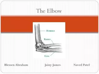
Disorders of The Elbow. Elbow Complex. Hinge type joint articulating between the humerus , radius & ulna Sites of Movement T rochlea of the humerus/trochlea of the ulna = ulnohumeral joint C apitulum of the humerus /head of the radius = radiocapitellar joint

E N D
Elbow Complex • Hinge type joint articulating between the humerus, radius & ulna • Sites of Movement • Trochleaof the humerus/trochlea of the ulna = ulnohumeraljoint • Capitulumof the humerus/head of the radius = radiocapitellarjoint • Proximal radioulnar joint (PRUJ)
Radius & Ulna Radius Head Neck Shaft Radial Tuberosity Interosseous Margin Ulna Coronoid Process UlnarTuberosity Trochlear Notch Head Interosseous Margin
Elbow Complex • Articulation and ligamentous anatomy • Humeroulnar Joint • Flexion and extension • Modified hinge joint • Humeroradial Joint • Flexion and extension • Pronation and supination • Modified ball & socket joint • Proximal and Distal Radioulnar joints • Pronation and supination • Pivot joint Compound Joint One articular capsule Three joints
Elbow Joint • Proximal Radioulnar Joint • Pivot joint - the radial head rotates against the radial notch of the ulna • The annular ligament of the radius holds the radial head in place • The joint capsule and synovial membrane is continuous with the elbow joint • Proximal Radioulnar Joint Ligament • Annular ligament that attaches to the ulna anterior & posterior to the radial notch forming a collar around the radial head, lined by a synovial membrane
Elbow Joint • Distal Radioulnar Joint (continued) • The disc separates the cavity of the distal radioulnar joint from the cavity of the wrist joint • A fibrous joint capsule surrounds the joint with an internal synovial membrane; there is a proximal extension of the synovial membrane forming a sacciform recess that serves to allow unimpeded movement of the radius about the stationary ulna
Elbow Joint • Distal Radioulnar Joint • Pivot type synovial joint – radius moves around the fixed distal end of the ulna • The head of the ulna articulate with the ulnar notch on the medial distal radius • A fibrocartilagenousarticular disc binds the ends of the radius and ulna • The base of the disc attaches to the medial edge of the ulnar notch and the apex of the disc is attached to the lateral side of the base of the ulnarstyloid process
Elbow Complex - Ligaments • Collateral ligaments are triangular bands formed by thickenings of the fibrous joint capsule • Medial – Ulnar Collateral Ligament (UCL) extends from the medial epicondyle to the coronoid process and olecranon. • Three parts: • 1. Anterior cord (the strongest) • 2. Posterior cord (fan-shaped, weakest) • 3. Oblique band • Lateral – Radial Collateral Ligament (RCL) from the lateral epicondyle of the humerus to blend with the annular ligament of the radius. This serves to hold the radial head in the radial notch of the ulna (allowing pronation/supination) • Interosseous membrane serves to transmit forces from the hand through the radius to the ulna then to the humerus
Joint Capsule • A fibrous joint capsule surrounds the joint and is attached to the humerus at the margins of the ends of the articular surfaces • A synovial membrane lines the capsule and is continuous with the proximal radio-ulnar joint • The capsule is strengthened by medial & lateral ligaments, the anterior and posterior aspects of the capsule are relatively weak
Bursae • OlecranonBursae: • Intratendinousolecranon bursa - sometimes present in the triceps tendon • Subtendonousolecranon bursa - between the olecranon and triceps tendon just proximal to the attachment to the olecranon • Subcutaneous olecranon bursa - located in the subcutaneous tissue over the olecranon
Muscles that Flex the Elbow • Biceps Brachii • Brachioradialis (work horse) • Brachialis
Muscles that extend the Elbow • Triceps brachii (medial head is work horse) • Anconeous
Muscles that Supinate the Forearm • Biceps Brachii (primary) • Supinator
Muscles that Pronate the Forearm • Pronatorquadratus (primary) • Pronatorteres
Structure Impacting ROM • Extension • Olecranonfossa, anterior capsule, biceps, brachialis • Flexion • Capsular tension • Supination • Anterior ligament, IOM • Pronation • Radial shaft, ulna
Muscle Attachments • Distal Shaft of Humerus • Brachialis, ECRL, ECRL, ECRB • Medial Epicondyle • PronatorTeres, PollicisLongus, FCR, FCU, FDS • Lateral Epicondyle • Anconeous, Brachioradialis, ECRL, ECRB, ECU, ED, EDM, Supinator
Distal Humerus Fractures • Intra-Articular Fractures • Deformity usually present • Elbow may dislocate • Presents like an avulsion fracture • Soft tissue attachments at the epicondyle are no longer functional
Type I Distal Humeral Fracture • Type I – Distal Humeral Fracture • Mechanism of Injury • Direct blow to partially flexed and pronatedelbow • Fall on partially flexed, abducted, and pronated forearm or on outstretched hand • Most common. Involves the majority of the capitellum and sometimes a small portion of the trochlea
Type II Distal Humeral Fracture • Type II Distal Humeral Fracture • Mechanism of Injury • Shear fracture of the articular surface with little or no subchondral bone damage • Occurs due to an indirect force being transmitted across the joint, i.e., hammering, lifting and similar activities
Type III Distal Humeral Fracture • Type III Distal Humeral Fracture • Intra-Articular fractures of the distal humerus • Comminuted compression or compression fracture • Often accompanied by a radial head fracture • Mechanism of Injury • Fall on outstretched hand
Extra-Articular Fractures of the Distal Humerus • Extra-Articular Fractures of the Humerus • Two Types: • Supracondylar fractures • Occurs just proximal to the humeral condyles • The most common of all distal humeral fractures • Transcondylar Fractures • Occurs across and through the humeral condyles • They are one-tenth as common as the supracondylar fractures
Extra-ArticularDistal Humeral Fractures • Extra-articular Distal Humeral Fracture • Mechanism Of Injury • Fall on outstretched hand • Can be regarded as both flexion and extension injuries • Clinical Evaluation • Angular deformity proximal to elbow • Rule Out vascular and nerve injury • Muscular contractions may be present
Clinical Evaluation of Distal Humeral Fractures • Little or no deformity • Progressive loss of motion • Palpable loose body in the antecubitalfossa • Crepitus with flexion and extension • Increased pain while moving elbow from extension to 45 degrees of flexion and rotating the forearm
Olecranon Fractures • Olecranon Fractures • Often occur as intra-articularfractures affecting stability of elbow • Fractures are displaced due to tendon and ligament attachments • Two forms of olecranonfractures • Non-displaced • Displaced
NondisplacedOlecranonFractures • NondisplacedOlecranon Fractures • Bone fragments separate no more than 2 mm and remain in contact during flexion and extension • Mechanism of Injury • Direct blow
Displaced OlecranonFractures • Displaced Olecranon Fractures • Four Types • Avulsion • Transverse • Comminuted • Fracture-dislocation • Mechanism of Injury • Fall on hand with elbow flexed and triceps contracting
OlecranonFractures • Avulsion – a transverse or oblique line dislodges a small non-articular portion of the olecranon, which is displaced by the pull of the triceps muscle insertion • Transverse or Oblique Fractures – fracture line will run from the deepest part of the semilunar notch transversely or obliquely to the crest of the ulna • Comminuted Fractures – fracture line runs in all directions. Some fragments may be extensively crushed. Distal humerus, radial head, and forearm fractures are commonly associated • Fracture Dislocations – fracture line is usually near the level of the tip the coronoid. Ulna will dislocate or subluxateanteriorly, along with the radius • Mechanism of Injury • Forceful direct blow to the posterior elbow
Clinical Evaluation OlecranonFractures • Nondisplaced fractures • Full ROM • Swelling and point tenderness over olecranon • Displaced fractures • inability to actively extend against gravity • point tenderness and swelling over the olecranon
Monteggia Fracture • Fracture of the ulna with dislocation of the radiocapitellararticulation • Most frequent in athletes • Four classifications
Monteggia Fracture • Type I • Anterior dislocation of the radial head associated with a fracture of the proximal ulna • The most common of these fractures • MOI • Direct blow to posterior ulna; fall on outstretched hand with forearm pronated • Clinical Evaluation • Pain, swelling, loss of elbow function • Forearm frozen in pronation • Anterior angulation of the fractured ulna
Monteggia Fracture • Type II • Posterior or posterlateral or dislocation of the radial head. Associated with a fracture of the proximal ulna • MOI • direct excessive rotational force • most common in middle aged women • Clinical Evaluation • posterior angulation of the fractured ulna • radial head can be palpated posteriorly • skin lesion will be posterior
Monteggia Fracture • Type III • Lateral or anterlateral dislocation of the radial head. Associated with a fracture of the proximal ulna • MOI • Direct blow on inner side of the elbow without rotation • Clinical Evaluation • Lateral angulation of the fractured ulna • Forearm frozen in midposition • Radial head can be palpated laterally
Monteggia Fracture • Type IV • An anterior dislocation of the radial head. Associated with a fracture of the proximal third of the radius and a fracture of the ulna at the same level • MOI • Fall on outstretched hand with forearm pronated • Mechanism can result in either type I or type II • Clinical Evaluation • anterior angulation of the fractured ulna • tenderness along radial shaft • radial head can be palpated in antecubitalfossa
Radial Head Fractures • Most common elbow fracture • Accompanied by tears and strains of surrounding ligamentous structures • Occurs more often in females • Four classifications • Type I: Non-displaced • Type II: Displaced • Type III: Comminuted • Type IV: Fracture dislocation
Radial Head Fracure • MOI • Falling on outstretched hand with elbow partially flexed and supinated • Can also occur by placing an excessive axial force on pronated forearm • Clinical Evaluation • Deformity • Type II only if fragment is displaced • Swelling • Displacement will cause soft tissue injury • Type III & IV commonly cause ligamentous or capsular disruption • Annular ligament, lateral collateral • Point tenderness over radial head
Elbow Dislocations • Second most common dislocation of a major joint • Associated with Fracture • Neurovascular assessment • Two classifications • Posterior • Anterior
Elbow Dislocations • Posterior dislocations – the most common of elbow dislocations. Position of forearm at onset determines whether it dislocates posterolateral or posteromedial • MOI • Fall on extended and abducted arm • Clinical Evaluation • Intense pain at the joint • Elbow flexed with an exaggerated prominence of the olecranon • Olecranon will be displaced for the plane of the epicondyles
Elbow Dislocations • Anterior dislocations – radius and ulna are displaced anterior to the distal humerus. Often accompanied by olecranon fracture • MOI • Strong blow to the posterior aspect of a flexed elbow • Clinical Evaluations • Elbow will be in full extension • Upper arm appears shortened, while forearm appears lengthened and held in supination
Arthritis of the Elbow • Two Forms • Traumatic • Non-traumatic • Both forms a capsular pattern of limitation • Cause must be determined • Patient history, functional evaluation, lab tests
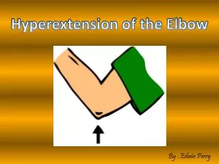
Traumatic Arthritis • Irritation of the synovial membrane as the result of trauma • Clinical Evaluation • Passive flexion is limited, elbow pain, MRI • MOI • Usually a result of hyperextension trauma, where the anterior capsule and the anterior part of the UCL are sprained
Nontraumatic Arthritis • Rheumatoid arthritis • Septic arthritis • Hematologic arthritis • Neurotrophic arthritis • Clinical evaluation • Athlete will c/o local pain, swelling and loss of motion • Lab tests
Clinical Evaluation of Elbow and Forearm • History • Onset (Acute versus Chronic) • Location • Referred pain to cervical spine, shoulder, hand • Mechanism of Injury • Technique • Associated sounds and sensations • Previous history • General Medical Health
Clinical Evaluation of Elbow and Forearm • Inspection • Contusion, ecchymosis, scars • Carry Angle (10-15 degrees) • CubitalFossa • Swelling • Medial Epicondyle • Flexor/Extensor muscle mass • Alignment of wrist forearm • Recurvatum
Clinical Evaluation of Elbow and Forearm • Palpation • Anterior • Biceps Brachii • CubitalFossa • Medial • UCL • Lateral • Radial Head • Capitulum • RCL • Annular ligament
Clinical Evaluation of Elbow and Forearm • Functional Testing • Elbow flexion/extension • Forearm pronation/supination • AROM • PROM • Ligamentous Stress Tests
Elbow Goniometry • Flexion/Extension • Patient • Standing, shoulder in neutral, forearm neutral • Fulcrum • Centered over olecranon • Stationary Arm • Aligned with long axis of humerus (use acromian as proximal landmark • Movement • Aligned with long axis of radius (use styloid as distal landmark)
Elbow Goniometry • Flexion/Extension • Patient • Supine, shoulder in neutral, forearm supinated • Fulcrum • Centered over olecranon • Stationary Arm • Aligned with long axis of humerus (use acromian as proximal landmark • Movement • Aligned with long axis of radius (use styloid as distal landmark)
Elbow Goniometry • Pronation/Supination • Patient • Humerus held against torso • Elbow flexed to 90 degree • Fulcrum • Centered to lateral ulnarstyloid process • Stationary Arm • Aligned parallel to midline of humerus • Movement