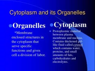
Cytoplasm
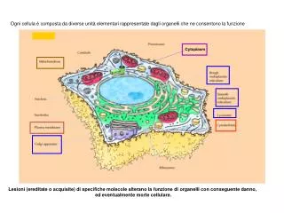
Ogni cellula è composta da diverse unità elementari rappresentate dagli organelli che ne consentono la funzione. Cytoplasm. Lesioni (ereditate o acquisite) di specifiche molecole alterano la funzione di organelli con conseguente danno, ed eventualmente morte cellulare.

Cytoplasm
E N D
Presentation Transcript
Ogni cellula è composta da diverse unità elementari rappresentate dagli organelli che ne consentono la funzione Cytoplasm Lesioni (ereditate o acquisite) di specifiche molecole alterano la funzione di organelli con conseguente danno, ed eventualmente morte cellulare.
PRINCIPALI ALTERAZIONI DELLA MEMBRANA PLASMATICA ALTERAZIONI GROSSOLANE DI STRUTTURA ALTERAZIONI DEL TRASPORTO ALTERAZIONI DI FUNZIONI RECETTORIALI
Fosfolipasi batteriche, rilasciate da cellule infiammatorie o rilasciate in seguito a pancreatiti possono alterare lipidi della membrana e causare lisi cellulare
Tossine batteriche inducono danno cellulare con diversi meccanismi 1. Membrana plasmatica (inducono alterazioni della permeabilità) 2. Componenti dell’apparato vescicolare (inibiscono fusione vescicole membrana plasmatica) 3. Componenti di vie di trasduzione del segnale (inibizione o attivazione della trasduzione del segnale) 4. Componenti del citoscheletro
Tossine batteriche inducono danno cellulare con diversi meccanismi 1. Membrana plasmatica (inducono alterazioni della permeabilità) 2. Componenti dell’apparato vescicolare (inibiscono fusione vescicole membrana plasmatica) 3. Componenti di vie di trasduzione del segnale (inibizione o attivazione della trasduzione del segnale) 4. Componenti del citoscheletro
1. Membrana plasmatica (inducono alterazioni della permeabilità) *Stafilococchi e Streptococchi rilasciano tossine (a-tossina e streptolisina O, rispettivamente ) che inducono la formazione di pori * Bacillus cereus ed altri rilasciano fosfolipasi e sfingomielinasi
2. Componenti dell’apparato vescicolare (inibiscono fusione vescicole membrana plasmatica) Neurotossine botuliniche e tetaniche determinano degradazione proteolitica di VAMP (vesicle-associated membrane protein, quale sinaptobrevina) o proteine “target” (T-SNARE, qualiSNAP-25, sintaxina)
Tossina tetanica BONT B,D,F,G BONT C BONT A,C,E
Un’esotossina della Bordetella pertussis ADP-ribosila e inattiva (mantiene in uno stato legato al GDP) la subunità ai delle proteine G trimeriche Le tossina colerica e l’enterotossina calore-sensibile di E. coli ADP-ribosilano e attivano (mantengono in uno stato legato al GTP) la subunità as delle proteine G trimeriche Tossine dei Clostridi ADP-ribosilano o glucosilano molecole della famiglia della Rho GTPasi (Rho, Rac, CdC42) inattivandole Una tossina della Yersinia pestis acetila in residui di Ser e Thr protein chinasi facenti parte della cascata delle MAP chinasi (MAP chinasi chinasi) e IKKb, inibendole e bloccando la cascata delle MAP chinasi e l’attivazione di NFkB 3. Componenti di vie di trasduzione del segnale (inibizione o attivazione della trasduzione del segnale)
Esotossine dei clostridi ADP-ribolisano G-actina disassemblando il citoscheletro. Componeneti superficiali si Shigella flexneri e Helicobacter pIlory inducono indirettamente polimerizzazione dell’actina, favorendo l’internalizzazione e la mobilità intracellulare del microrganismo. 4. Componenti del citoscheletro
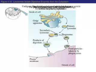
Salmonelle e Shigelle traslocano proteine effettrici nella cellula ospite stimolando la formazione di ruffles di membrana che internalizzano il battere Shigella, Listeria e Rickettsia escono dal vacuolo e inducono la formazione actina fila- mentosa. Legionella e Brucella inducono la formazione di un vacuolo con diverse membrane e si repli- cano nel REL Mycobacterium e Salmonella bloccano la ma- turazione del vacuolo di fagocitosi
Figura 2.25 - Schema degli scambi attraverso le membrane biologiche. Dal volume: Pontieri “Patologia Generale” Piccin Nuova Libraria S.p.A.
Da: Harrison’sPrinciple of Internal Medicine; Chapter 365. InheritedDefects of Membrane Transport
“CANALOPATIE” Kass. J. Clin. Invest. 115, 1986-1989, 2005
Podosomes are circularstructuresformingprotrusions of the ventral membrane containingintegrins, cytoskeletalproteins, tyrosinekinases and tyrosinephosphorylatedproteins The distribution of podosomes in osteoclasts cultured on bone laminae: Effect of retinol Alberta Zambonin‐Zallone, Anna Teti, Aldo Carano, Pier Carlo Marchisio Abstract: Osteoclasts, isolated and purified from the medullary bone of calcium‐deficient egg‐laying hens, adhere to glass coverslips in vitro by means of specialized protrusions of the ventral membrane, denoted podosomes. These structures represent dotlike close‐contact adhesion sites in which most cytoskeletal proteins involved in linking the plasma membrane to microfilaments are organized according to a specific and previously described pattern also shared by many oncogene‐transformed cells. We show nowthatpodosomes are notonly a feature of osteoclastsadheringtoartificialglasssurfacesbut are alsopresent in the ventral membrane of osteoclastsadheringtobonelaminae. Moreover, the quantity and the topography of podosomes may be modulated by retinol, which increases bone‐resorbing activity of osteoclasts both in vivo and in vitro. A comparative transmission electron microscopystudy of osteoclastsadhering on bonelaminae in vitro or in vivo indicatesthatpodosomeswithidenticalfeatures are present in the clear zone of the osteoclasts in eithercondition. Sincepodosomes are the sealingstructures of the clear zone, podosomeformationmayrepresentone of the modificationsinvolved in the reorganizationprocess of the osteoclastthatprecedesboneresorption. J. Bone and MinMetab. 3:517, 1988 Cell. Signal. 2011
Receptor for Activation of Nuclear factor Kappa B/Ligand 1,25 (OH)2 D3 Osteoprotegerin
“Canalopatie” Kass. J. Clin. Invest. 115, 1986-1989, 2005 Episodi di tachicardia ventricolare durante il rilascio di catecolamine in seguito a stress emozionali o fisici
Sensore di voltaggio nel muscolo scheletrico Recettore RYR1 (Rianodina) Ca2+ Store Intracell Reticolo Sarcoplamico Meccanismi di regolazione della liberazione di calcio nel muscolo scheletrico e cardiaco Canale per il Ca2+ (VOC) nel muscolo cardiaco Ca2+ Recettore RYR2 Ca2+ “Calcium-inducedcalcium release” (CICR)
Figura 2.32 - Struttura dei trasportatori a 12 domini tansmembranacei e loro variante ABC (ATP-Binding Cassette). Dal volume: Pontieri “Patologia Generale” Piccin Nuova Libraria S.p.A.
MALATTIE CAUSATE DA ALTERAZIONI DI GENI CODIFICANTI PER TRASPORTATORI ABC (“ATP-BINDING CASSETTE”) Uitto J, TRENDS in Mol Med 11, 341-343, 2005
LESIONI GENE ABCA1 E ABCG5/8 COMPORTANO ECCESSIVO ACCUMULO DI COLESTEROLO NEI TESSUTI
intestino chilomicrone tessuti periferici microcircolo lipoproteinlipasi fegato VLDL IDL LDL
48 100 100
DNA Gene per APO B-100 2153 mRNA CAA glutamina deaminasi intestinale UAA Codone di stop APOB-100 APOB-48
ESPRESSIONE E FUNZIONI APOPROTEINE APOPROTEINAESPRESSIONE FUNZIONE APO-B100VLDL-IDL-LDLLigando per Recettore APO-B48Chilomicroni Ligando per Recettore APO-AHDLAttivatore di Enzima (aciltransferasi) APO-CVLDL-IDLAttivatore di Enzima Chilomicroni(lipoprotein lipasi) APO-EVLDL-IDL-HDLLigando per Recettore Chilomocroni APO(a)VLDL-IDL-LDL Inibitore Fibrinolisi
epatocita tessuti periferici HDL High Density Lipoprotein TRIGLICERIDI ( 2%) COLESTEROLO (18%) FOSFOLIPIDI (30%) PROTEINE (50%) a-HDL APO-E APO-A APO-A pre-b-HDL APO-E FOSFOLIPIDI Colesterolo libero intestino PLTP: phospholipid transfer protein Tessuti periferici (e macrofagi)
fegato LCAT Lecitina Colesterolo Acil Transferasi APO-A CETP HDL APO-A a-HDL pre-b-HDL APO-A APO-E VLDL/IDL LDL Tessuti periferici PLTP CETP* *Colesteryl Esther Transfer Protein
Lipoprotein metabolism has a key role in atherogenesis. It involves the transport of lipids, particularly cholesterol and triglycerides, in the blood. The intestine absorbs dietary fat and packages it into chylomicrons (large triglyceride-rich lipoproteins), which are transported to peripheral tissues through the blood. In muscle and adipose tissues, the enzyme lipoprotein lipase breaks down chylomicrons, and fatty acids enter these tissues. The chylomicron remnants are subsequently taken up by the liver. The liver loads lipids onto apoB and secretes very-low-density lipoproteins (VLDLs), which undergo lipolysis by lipoprotein lipase to form low-density lipoproteins (LDLs). LDLs are then taken up by the liver through binding to the LDL receptor (LDLR), as well as through other pathways. By contrast, high-density lipoproteins (HDLs) are generated by the intestine and the liver through the secretion of lipid-free apoA-I. ApoA-I then recruits cholesterol from these organs through the actions of the transporter ABCA1, forming nascent HDLs, and this protects apoA-I from being rapidly degraded in the kidneys. In the peripheral tissues, nascent HDLs promote the efflux of cholesterol from tissues, including from macrophages, through the actions of ABCA1. Mature HDLs also promote this efflux but through the actions of ABCG1. (In macrophages, the nuclear receptor LXR upregulates the production of both ABCA1 and ABCG1.) The free (unesterified) cholesterol in nascent HDLs is esterified to cholesteryl ester by the enzyme lecithin cholesterol acyltransferase (LCAT), creating mature HDLs. The cholesterol in HDLs is returned to the liver both directly, through uptake by the receptor SR-BI, and indirectly, by transfer to LDLs and VLDLs through the cholesteryl ester transfer protein (CETP). The lipid content of HDLs is altered by the enzymes hepatic lipase and endothelial lipase and by the transfer proteins CETP and phospholipid transfer protein (PLTP), affecting HDL catabolism.
LUME INTESTINO ABCG5/ABCG8 (Sitosterolemia) LUME CANALICOLO BILIARE ABCG5/ABCG8 (Sitosterolemia)
Trasportatori steroli vegetali PS: steroli vegetali MTP: microsomal transfer protrein
J. Couzin-Frankel Science 324, 1504 -1507 (2009) Published by AAAS
J. Couzin-Frankel Science 324, 1504 -1507 (2009) Published by AAAS
Regulatory Nucleotide Binding Domain
Figura 2.37 - Le 4 classi delle principali mutazioni del trasportatore ABC. IV Dal volume: Pontieri “Patologia Generale” Piccin Nuova Libraria S.p.A.
CFTR mutata induce infiammazione polmonare Cuore polmonare
3 PMN 2a IL-8 ED ALTRE CITOCHINE 2b INIBIZIONE DI AUTOFAGIA CFTR MUTATA ACCUMULO DI CERAMIDE PMN Aumentata morte cellulare Rilascio di DNA IL-8 ED ALTRE CITOCHINE PMN NETs*** 4 Riduzione battito ciliare e lavaggio vie aeree PMN Muco denso e viscoso Stimolazione CXCR2 INFEZIONE (P. aeruginosa) 1 PMN + “shedding CXCR1** RIDUZIONEASL* CFTR MUTATA ** recettore per IL-8 che attiva capacità microbicide verso P. aeruginosa *ASL = AIRWAY SURFACE LIQUID *** Neutrophil extracellular traps