Download

1 / 15
150 likes | 253 Views
Understanding key points of DNA sequencing: DNA fragments, radioactivity, electrophoresis, autoradiography. Learn the process & interpret data. Helpful insights for biology students.

E N D
DNA SEQUENCING Module BYB2 KGV Biology Cliff Sharp
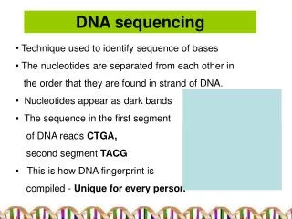
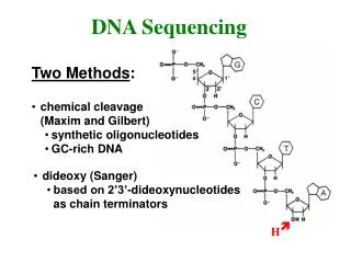
This is a technique used to find the sequence of bases in a DNA sample. It is quite complicated and you DO NOT have to learn it all. There are different methods of doing this, but they will be described in questions and you will have to interpret the information. KGV Biology
This one method. DO NOT LEARN IT! The main points are:- The length of the DNA fragments The radioactivity involved Electrophoresis Autoradiography Understand those parts!!! KGV Biology
This is the segment of DNA we wish to sequence. AGTCATCGG It is copied by PCR. AGTCATCGG AGTCATCGG AGTCATCGG AGTCATCGG AGTCATCGG AGTCATCGG AGTCATCGG AGTCATCGG KGV Biology
A small quantity of chemically modified nucleotide with the base T is made radioactive. It is added to a mixture of nucleotides with the bases A, T ,G and C. Some of our DNA fragments are added to the mixture and an enzyme is added which starts DNA replication. The process stops when the modified nucleotide with the radioactive T is included in the copied fragment, like this :- AGTCATCGG AGT AGTCAT KGV Biology
The same is done with the other 3 bases (in separate tubes!) For G it looks like this:- AGTCATCGG AG AGTCATCG AGTCATCGG For C it looks like this:- AGTCATCGG AGTC AGTCATC And for A it looks like this :- AGTCATCGG AGTCA A KGV Biology
The different sized fragments are then transferred to wells in one end of a gel block. The differently labelled fragments are kept separate, The radioactive T in one well, C in another and so on. Afragments T fragments G fragments C fragments KGV Biology
This is the bit you do need to learn!! ELECTROPHORESIS This is used to separate pieces of DNA according to their MASS or DENSITY Once the fragments are placed in in the gel an electric current is applied. Afragments T fragments G fragments C fragments KGV Biology
A AG AGT AGTC AGTCA AGTCAT AGTCATC AGTCATCG AGTCATCGG A T G C Electric current applied KGV Biology
You will notice that the smaller fragments (those with a lower mass or density) travel FURTHER in the gel. The longer fragments (with higher mass/more dense) do not travel far. KGV Biology
The gel is placed close to photographic film. (Never write just “film”. Say what type of film.) A The film is then developed. AG The radioactivity “fogs” the film causing dark bands to appear. This is called an autoradiogram. AGT AGTC AGTCA AGTCAT AGTCATC AGTCATCG AGTCATCGG KGV Biology
You now have a developed piece of film looking like this. KGV Biology
You know which column held each base and you can see the relative distances travelled. You can now work out the base sequence. (Read from the bottom upwards). A T G C KGV Biology
A T G C So what is this base sequence? GACTGCATA KGV Biology
A similar method is to isolate the DNA and cutting it into smaller pieces using restriction endonuclease. The fragments can then be multiplied using PCR. Radioactive probes may then be used. These are single-stranded sections of DNA which will attach to the complementary base sequences on the DNA being investigated. Electrophoresis then autoradiography are then carried out. KGV Biology