Download
1 / 33
330 likes | 347 Views
Explore the composition and roles of erythrocytes, leukocytes, platelets in the blood, and their functions in physiological processes like oxygen transport, immune defense, and clotting. Learn about erythropoiesis, leukocyte types, and hemostasis mechanisms.

E N D
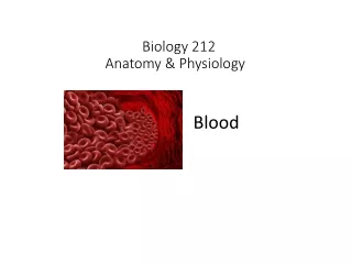
Blood: Volume: 4 - 6 liters, ~ 8% to 9% of body weight Components: Formed Elements: Erythrocytes (Red blood cells) Leukocytes (White blood cells) Platelets Plasma: Water Many proteins Ions, nutrients, wastes, hormones, etc pH: 7.35 - 7.45, Buffered to remain stable
Erythrocytes Primary Function - Transport of oxygen and carbon dioxide Cytoplasm full of hemoglobin Biconcave disks ~7.5 um diameter ~2 um thick No nucleus 4.5-5.5 million per microliter (cubic millimeter) 4.5-5.5 billion per milliliter (cubic centimeter) Slightly higher in men
Hemoglobin Each molecule: 4 large Globin proteins Each surrounding an iron- containing Heme Group Oxygen reacts reversibly with iron: Binds in lungs Unbinds in consumer tissues
Erythrocyte formation = erythropoiesis Occurs in bone marrow Developing erythrocytes = erythroblasts
Erythropoiesis regulated by hormone erythropoietin produced by kidneys
Erythrocytes also carry specific glycoproteins, or antigens, on their surfaces which are responsible for blood types If transfused into person with incompatible blood type, antibodies in recipient's serum will react with these antigens & cause erythrocytes to stick together or agglutinate
Erythrocytes normally survive ~120 days in circulation Trapped and destroyed (mostly in spleen) Iron salvaged from hemoglobin, returned to bone marrow
Leukocytes (White blood cells) Five different types of cells All formed in bone marrow 4,000 to 10,000 per cubic millimeter or 4,000,000 to 10,000,000 per milliliter (cubic centimeter)
Each type has specific functions, but in general: Leukocytes function in body defenses by: Engulfing & digesting invading organisms & debris Directly killing invading cells Producing antibodies Secreting chemicals which activate other immune cells Secreting chemicals which promote inflammation
Leukocytes Grouped into two categories based upon appearance of granules in cytoplasm: Abundant, distinct granules = Granular Leukocytes or Granulocytes Few, small granules = Agranular Leukocytes or Agranulocytes
Granular Leukocytes Named according to how these granules react to routine lab stains ("Wright's Stain" is most common) The nucleus of each type also has a characteristic shape and/or density Three types: Neutrophils, Eosinophils, Basophils
Neutrophils 40% - 60% of leukocytes in blood. Granules present in cytoplasm but stain weakly with both acidic and basic stains Nucleus has 3 to 7 lobes Also called Polymorphonuclear Leukocytes
Eosinophils 1% - 4% of leukocytes in blood. Granules attract acidic stain eosin, therefore stain red or orange Nucleus is indented or bilobed
Basophils 0.5% - 1% of leukocytes in blood. Granules attract the basic stain hematoxylin, therefore stain blue or purple Nucleus is indented or bilobed, but is often obscured by the granules Outside of circulation: Mast cells
Agranular Leukocytes Two unrelated types of leukocytes, neither of which has abundant granules: Lymphocytes, Monocytes The nucleus of each type also has a characteristic shape and density
Monocytes 4% to 8% of leukocytes in blood Nucleus indented, may be kidney shaped or horseshoe shaped. Stains only moderately blue. Cytoplasm is abundant, typically stains an even light blue or bluish-gray Outside of circulation: Macrophages
Lymphocytes 20% to 40% of leukocytes in blood Nucleus usually spherical, may be slightly indented. Stains darker blue. Varying amounts of cytoplasm which stains a pale to medium blue
Lymphocytes Two types of lymphocytes with different functions in immune system, but they appear identical in blood: T-lymphocytes and B-lymphocytes
All leukocytes formed in bone marrow, then enter blood But: Not particularly active when in the blood. Most leukocytes are using the blood to get to other tissues and organs, where they differentiate and become active
Since they generally function outside of the circulatory system, primarily in the connective tissues of other organs, All leukocytes can leave (and most can also reenter) the blood vessels by a process called Diapedesis. Therefore: All of the leukocytes, and the cells which they mature into, are normally found in connective tissues throughout the body
Platelets Function in clotting of blood when vessels are injured 250,000 to 500,000 per cubic millimeter or 250,000,000 to 500,000,000 per milliliter (cubic centimeter)
Platelets Fragments of much larger cells, called megakaryocytes, which remain in the bone marrow
When blood vessels are damaged, the flow of blood through them must be stopped until the body can repair the injury. This is called Hemostasis It involves three processes in rapid sequence: Narrowing of the vessel reduces the flow of blood near the injury Vasospasm
When blood vessels are damaged, the flow of blood through them must be stopped until the body can repair the injury. This is called Hemostasis It involves three processes in rapid sequence: Vasospasm Platelet Plug
Formation of a Platelet Plug - Under normal conditions, platelets do not stick to each other or to the walls of blood vessels - When a blood vessel is injured, it releases chemicals which cause platelets to attach to each other and to the injured part of the vessel. This is platelet activation. - As new platelets attach, they also become activated and release more chemicals which attract and activate more platelets
When blood vessels are damaged, the flow of blood through them must be stopped until the body can repair the injury. This is called Hemostasis It involves three processes in rapid sequence: Vasospasm Platelet Plug Fibrin Clot
Formation of a Fibrin Blood Clot - This involves a series of sequential chemical reactions in which the products of the first reaction serve as the catalysts of the second reaction, whose products are the catalysts of the third reaction, whose products are the catalysts of the fourth reaction, etc. - This allows for a very rapid increase in the rate at which the clot forms. - It also allows many chances to stop the process if it began by mistake or gets too far away from where the vessel was injured.
Formation of a fibrin blood clot involves 13 "clotting factors". Each of these is the reactant for the next of these sequential reactions
Formation of a fibrin blood clot involves 13 "clotting factors". Each of these is the reactant for the next of these sequential reactions Last reaction: Plasma protein fibrinogen is converted to large fibers of fibrin, which forms a dense mesh and traps erythrocytes, platelets, and leukocytes
Once the fibrin clot has formed, it is stabilized by enzymatic formation of covalent crosslinks among the strands of fibrin. Over the next 30 to 60 minutes, the clot retracts, becoming denser and more solid. End result: Dense network of interconnected strands of fibrin with platelets, erythrocytes, and leukocytes trapped within it.
Over next few days, after vessel has time to repair itself: Clot is dissolved and removed by process called fibrinolysis. Plasma protein plasminogen converted to active enzyme plasmin which dissolves fibrin.