Download
1 / 16
160 likes | 230 Views
This study examines cervico-vaginal infections in female patients aged 14-16 years in Lagos, Nigeria, identifying predominant microorganisms and their antibiotic susceptibility patterns. It highlights the health implications of the identified organisms and emphasizes the importance of proper antibiotic use. The research underscores the significance of maintaining a balanced vaginal ecosystem to prevent infections and associated complications.

E N D
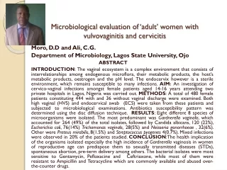
Microbiological evaluation of ‘adult’ women with vulvovaginitis and cervicitis Moro, D.D and Ali, C.G. Department of Microbiology, Lagos State University, Ojo ABSTRACT INTRODUCTION: The vaginal ecosystem is a complex environment that consists of interrelationships among endogenous microflora, their metabolic products, the host’s metabolic products, oestrogen and the pH level. The endocervix however is a sterile environment, which remains susceptible to many infections. AIM: An investigation of cervico-vaginal infections amongst female patients aged 14-16 years attending two private hospitals in Lagos, Nigeria was carried out. METHODS: A total of 480 female patients constituting 444 with and 36 without vaginal discharge were examined. Both high vaginal (HVS) and endocervical swab (ECS) were taken from these patients and subjected to microbiological examinations. Antibiotics susceptibility pattern was determined using the disc diffusion technique. RESULTS: Eight different 8 species of microorganisms were isolated. The most predominant was Gardnerellavaginalis, which accounted for 264 (49%) of the total isolates, followed by Candida albicans, 120 (22%), Escherichia coli, 76(14%) Trichomonasvaginals, 28(5%) and Neisseriagonorrhoeae , 32(6%). Other were Proteus mirabilis, 8(1.5%) and Streptococcus pyogenes 4(0.7%). Mixed infections were observed in 20% of the patients studied. CONCLUSION:The health implication of the organisms isolated especially the high incidence ofGardnerellavaginosis in women of reproductive age can predispose them to sexually transmitted diseases (STDs), spontaneous abortion, pre-term delivery among others. The bacterial isolates were most sensitive to Gentamycin, Pefloxacine and Ceftriaxone, while most of them were resistant to Ampicillin and Tetracycline which are commonly available and abused over-the-counter drugs.
INTRODUCTION • Vaginal ecosystem, a complex with inter relationships among endogenous. • In addition, metabolic products of the microflora and host, gestrongen and low PH (ACOG, 1996) • Dynamic equilibrium of vagina challenged by endogenous and exogenous factors. • Endocerviis lined up with columnar epithelium replaced by division of basal cells. • Endocervix is a naturally sterile environment, but susceptible to many infectious (Jenny, 1990)
Vaginitis and cervicitis, reproductive tract infections (RTI), are frequent and troublesome disorders in women/s reproductive life. • Vaginitis and cervicitis constitute great socioeconomic and medical problems. • Both often trivialized by medical practitioners but are extremely common with considerable distress and suffering (Sobel, 1990). • Normal pH level of vagina is 3.5 – 4.2 stabilizes this ecosystem • Alteration of vaginal ecosystem due to medication repeated douching, self diagnosis or self treatment (March, 1991). • Other such factors are antibiotics, hormones, contraceptives vaginal mediation sexual intercourse, STDs, stress etc/Martens • Several bacteria and other microbes have been implicated in vulvovagnitis and cervicitis (Adinmaet al 2000).
MATERIALS AND METHODS Study Area and Subjects • This study was earned out in Jim-Sam hospital and maternity home, Ijora – Badia, Lagos for 2 years, 2014 – 2016. • Patients were interviewd before specimen collection. • 480 female patients, 444 symptomatic and 36 without vaginal discharge. • A questionnaire was specially designed and used.
Sample Collection and Analysis • On examination bed, external vaginal surfaces examined for required signs. • Two high vaginal swab (HVS) and two endocervical swab (ECS) obtained. • pH of discharge don with a strip of indicator paper. • Discharges collected in the lower speculum
Microbiological Analysis • Swab specimens divided into two (i) Culture on selective media (ii) For direct staining and microscopy • Media: MacConkey agar, Chocolate agar, Blood agar, Sabour and dextrose agar and Thayer Matin agar • Microorganisms identified after 18 – 24 h incubation morphological cultural and biochemical characteristics • Antibiotics susceptibility was carried out using Kirby Bauer method (Bauer et al, 1966). • Whiff test or amine test for yeast cells, due cells. • Wet mount: clue cells, budding yeasts motile bacteria trichomonads and PMN cells (pus cells) (Cheesbrough, 2002).
RESULTS Table 1: Age distribution of women examined Table 1 shows the age distribution of women examined. Range 14 – 63 years • 88.3% of the subjects were 40 years and below (Table 1) • Vulvovaginitis and cervicitis reduced with age.
Table 2: Marital status of women studied more than More than half of the women practice monogamy and one quarter practice polygamy (Table 2)
Table 3: Distribution of women on contraceptive use • About half of the subjects do not practice any form of contraceptive while condom and coil were the most usually practiced contraceptive methods (Tables 3)
Table 4: Organisms isolated and sites of isolation • Table 4 shows thatG.vaginalis, C. albicansandE.coliwere the most implicated isolates in vaginitis and cervicitis.
Table 5: Distribution of microbes in the sample analyzed • Single and mixed infections were found. Single infections constituted 71.6% and only 17.6% were mixed. (Table 5)
Table 6: Antibiotics susceptibility pattern of bacterial isolates • Gentamycin, nitrofurantoin, perfloxacin and ceftriaxones showed high in – vitro efficacy against most isolates testd while the commonly used antibiotics were resisted by the isolates. (Table 6)
DISCUSSION • Majority of the infected women fell between 10 and 40 years, sexually active age group • This shows that sexual intercourse may predispose women to infection • Women who practice monogamy are likely to have more frequent sexual intercourse (Mattson et al., 2016) • This may likely be responsible for the high incidence of vulvovaginitis and cervicitis (Williams et al; 2004) • The 36 control who cut across the various marital status showed no evidence of infection, so these infectious are highly symptomatic • Contraception appears to be associated with these infections as those who did not use any harboured no pathogen.
DISCUSSION CONTD. • Those who use condom, coil and IUCD were more infected. • Single infections were more predominant as combined/mixed infections were fever. • Microbial pathogens were not recovered from the asymptomatic subjects • Bacterial high susceptibility to gentamycin, nitrofurantoin, perfloxacin and ceftriaxone. • However, a high resistance to commonly used antibiotic may be due to abuse.
REFERENCES • ACOG technical bulletin (1996): Vaginitis Internet. J GynaccolObstets. 54: 293 – 302. • Adekunle, O.A and Ladipo, O.A (1996). Reproductive tract infections in Nigeria: challenges for a fragile health infrastructure In German, A., Holmes, K., Piot P. and Wassartier A. (Eds) Textbook on Reproductive tract infection. Plenum Press New York, pp 297 – 316. • Adinma, J. I. B., Okiwoli, N.R., Agbai, A.O. and Inaeze, N.C. (2000) Gardnerellavagnalis: A comparison of two methods of laboratory detection Nig. Qt. J. Hosp. Med. 10 (1): 38 – 40.
Cheesbrough, M. (2001) Identification of Micro - organisms: In medical laboratory manual for tropical countries Vol. II pp 252 – 280. • Govender, L., Hoosen, A.A., Moodley, J., and Moodley, P. (1996). Bacterial vaginosis and associated infections in pregnancy Intern. J. Gynaeccol. Obstet. 55:23 – 28. • Jenny , C., Hooten T.M, Bowers, A. et al. (1990) sexually transmitted diseases in victims of rape. N. Eng. J. Med. 322: 713 – 716. • Sobel J.D. (1990) Vaginal infections in adult women, Med. Clin. North Am. 74:1573 – 1601. Williams , M.D., Smith, J.J., Soble J.J., et al (2004) sexually transmitted disease J. Urol. Channel 20:25 – 30.