Understanding Digestion: The Key to Nutrition and Energy
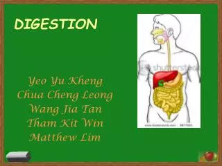
Digestion is a vital process that transforms the food we eat into usable nutrients for our body. It involves the mechanical and chemical breakdown of ingested food into smaller molecules like amino acids and glucose that can be absorbed into the bloodstream. This process begins in the mouth with saliva and continues through the esophagus, stomach, and intestines, ultimately allowing the body to utilize these nutrients for energy and cell maintenance. Proper digestion ensures adequate nutrition, health, and the elimination of waste.

Understanding Digestion: The Key to Nutrition and Energy
E N D
Presentation Transcript
Why is digestion important? When you eat foods—such as bread, meat, and vegetables—they are not in a form that the body can use as nourishment. Food and drink must be changed into smaller molecules of nutrients before they can be absorbed into the blood and carried to cells throughout the body. Digestion is the process by which food and drink are broken down into their smallest parts so the body can use them to build and nourish cells and to provide energy
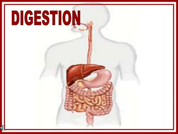
Introduction The body uses a variety of small molecules (amino acids, fatty acids, glucose) for its metabolic needs. Food is mechanically and chemically broken down into these molecules during digestion, after which they can be taken up by body cells through the separate process of absorption. Food travels in a one-way path from mouth to esophagus to stomach to small intestine to large intestine to anus. Organs and structures in the digestive system are specialized for specific functions in digestion. Digestive enzymes are specific hydrolytic enzymes that have a preferred temperature and pH. Proper nutrition is necessary to health.
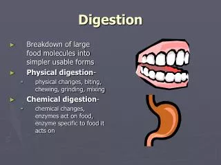
Basic Functions Digestion: the mechanical and chemical breaking down of ingested food into particles, then into molecules small enough to move through epithelial cells and into the internal environment. Absorption: the passage of digested nutrients from the gut lumen into the blood or lymph, which distributes them through the body. Elimination: the expulsion of indigestible residues from the body.
DIGESTION During digestion, proteins are broken down into ______, carbohydrates into _______, fat to ______, and nucleic acids to _______. Amino acids, glucose, glycerol/fatty acids, nucleotides. Digestion is an extracellular process. It occurs within the gut (a tube that runs from mouth to anus).
Step 1 of Digestion When you chew food, you moisten and lubricate it with saliva. Saliva contains water, mucus, and salivary amylase, a hydrolytic enzyme that breaks down starch in the presence of water. Starch is broken down to maltose (a disaccharide of glucose), which is later broken down to glucose in the intestine. Thus, digestion begins in the mouth, even before the food is swallowed. Once food has been chewed, it is called a bolus. Food is then passed through the back of the mouth when you swallow. The first region that it enters is called the pharynx, which is simply the region between mouth and esophagus where swallowing takes place. Swallowing is a reflex action (requires no conscious thought).
ESOPHAGUS a long muscular tube that extends from pharynx to stomach. The inner surface lined with mucus membranes. This layer is attached by connective tissue to a layer of smooth muscle containing both circular and longitudinal muscle. food moves down the esophagus through Peristalsis (rhythmical contractions of the esophageal muscles). Food bolus reaches the end of the esophagus and arrives at the cardiac sphincter connecting to the stomach. (sphincters function like valves. Made of muscles that encircle tubes. The tubes open them when the sphincters relax, and close them when they contract). Normally, this sphincter prevents food from moving up out of stomach, but when vomiting occurs, a reverse peristaltic wave causes the sphincter to relax and the contents of the stomach are propelled outward.
Esophagus and stomach http://www.metacafe.com/watch/6336127/gastroscopy/ Gastroscopy
STOMACH Thick-walled, organ that lies on left side of the body beneath the diaphragm. can stretch to hold about 2 liters of solids/liquids in an average adult. three layers of muscle contract to churn and mix its contents “hunger pains” are felt when an empty stomach churns. the mucus lining of the stomach contains inner Gastric Glands which produce and release gastric juice. Gastric juice contains pepsinogen and HCl (hydrochloric acid). HCl converts pepsinogen to pepsin by removing part of the molecule, exposing the molecule’s active site. Pepsin, a hydrolytic enzyme that breaks down proteins into smaller chains of amino acids called peptides. (further on in the digestive tract they are broken down individual amino acids by other enzymes.
Stomach continued HCl gives stomach a pH of 2~3. Highly corrosive. This kills bacteria in food and helps break it down The stomach is protected by a thick layer of mucus secreted by epithelial (epithelium is the lining of the stomach) cells to help protect the stomach wall from being digested. Still the epithelium is constantly eroded and mitosis completely replaces it every three days. If HCl does penetrate the mucus, pepsin starts to digest the stomach lining ---> forms an ulcer however, the #1 cause of ulcers is actually a bacterial infection (Helicobacter pylori) that impairs the ability of cells to produce mucus. Thus, most ulcers can now be cured with antibiotics. after 2 - 6 hours (depending on the type of food), the food has been turned into a semi-liquid food mass called acid chyme, and the stomach empties into the first part of the small intestine (called the duodenum). This emptying is controlled by the pyloric sphincter at the bottom of the stomach.
Small Intestines: the food processor Only some digestion has thus far taken place. Most of digestion and absorption of most nutrients occur in the small intestine. Divided into three zones: the duodenum, jejunum, and ilium. is about 6 meters long (~20 feet), compared to 1.5 m (~ 5 feet) for large intestine. first 25 cm of small intestine called the Duodenum. The duodenum plays a major role in digestion. It is here that secretions sent from the liver and pancreas break down fat and peptides, and secretions of the duodenum itself also break down other nutrients.
Small Intestines continued first 25 cm of small intestine called the Duodenum. The duodenum plays a major role in digestion. It is here that secretions sent from the liver and pancreas break down fat and peptides, and secretions of the duodenum itself also break down other nutrients. walls of the duodenum are lined with millions of interstitial glands that produce juices containing enzymes that finish the digestion of protein and starch. secretions from the interstitial glands contain digestive enzymes: peptidases digest peptides to amino acids. Disacharides brush against the wall of the duodenum where different enzymes continue work on the different disaccharides maltase digests maltose (a disaccharide) to glucose. Other enzymes made here digest other disaccharides (e.g. lactase digests lactose, the sugar in milk). This happens right next to the wall of the intestine so that it is easily absorbed.
The structure of the small intestine is well related to its function of absorption. 1. Long with convoluted walls to increase surface area 2. Surface area further increased by presence of finger-like projections called villi (a single one is called a “villus”. Interstitial glands are at the base of each villi. 3. Villi themselves are lined with columnar cells coated with microvilli. Each villi contains blood vessels and lymph vessels (lacteal). absorption takes place across the wall of each villus ---> this can happen passively or actively. Recall that active transport across cell membranes requires ATP. The nutrient can now enter the blood or the lymphatic system, depending on what type it is.
continued Fatty acids and glycerol are absorbed across the villi, are recombined into fat molecules in the epithelial cells of the villus. The fats then move into the lacteal of each villus and enter the lymphatic system. sugars and amino acids enter the blood through the capillary network. The blood vessels from the villi in the small intestine merge to form the hepatic portal vein which leads to the liver. http://www.youtube.com/watch?v=AJ1wKsmBPvA
Liver The liver is a major player in the overall function of the circulatory system. The liver is often likened to a checkpoint. Substances that enter the body through the small intestine are first screened by the liver. If those substances are found to be beneficial, they are allowed to circulate to the rest of the body's organs. But if those substances are harmful, the liver is able to isolate and rid the body of them before they damage other internal organs.
Liver the Liver produces bile, which is sent to the duodenum via a duct from the gall bladder (where bile is stored). Bile is a thick green liquid (it gets its green colour from by-products of hemoglobin breakdown (another function of the liver). Bile contains emulsifying agents called bile salts which break fat into fat droplets.
Functions of the Liver 1. keeps blood concentrations of nutrients, hormones, and sugar etc. constant (e.g. converts glucose to glycogen and back to keep blood glucose levels constant). 2. Interconversions of nutrients (e.g. carbohydrates to fats, amino acids to carbohydrates and fats). 3. removes toxins from the blood (detoxifies). Removes unwanted particulate matter from the blood through the mediation of macrophages. 4. Production of Bile. Up to 1.5 liters of bile per day! 5. Destroys old red blood cells. 6. Production of urea.
Continued. 7. Manufacture of plasma proteins such as fibrinogen and albumin. 8. Manufacture of cholesterol. 9. Storage of iron. 10. Storage of vitamins. 11. In embryos (of vertebrates) , the liver makes Red Blood Cells
Pancreas Pancreas sends pancreatic juice into duodenum through duct the juice contains enzymes and sodium bicarbonate (NaHCO3) NaHCO3 makes the juice highly alkaline (pH ~ 8.5). It neutralizes the acid chyme and make the small intestine pH basic pancreatic juice contains hydrolytic enzymes including pancreatic amylase (digests starch to maltose), trypsin (digests protein to peptides), and lipase (digests fat droplets to glycerol & fatty acids).
metabolism and uptake by the body. As the glucose passes through the walls of the intestines (upper left) to the bloodstream, a signal is sent to the pancreas (yellow, upper centre) to secrete the hormone insulin (green strands in blood vessel). Insulin converts glucose into its storage form, glycogen, which is then stored in the liver (centre), muscle cells (lower left) and adipose (fat) cells (lower right) until it is needed for energy.
Large Intestines consists of colon and rectum (the rectum is the last 20 cm of the colon). Opening of rectum is called anus. colon has 3 parts (ascending, transverse, and descending)
LARGE INTESTINE reabsorption of water from indigestible food matter (feces) absorption of certain vitamins feces also contains bile pigments, heavy metals, and billions of E. coli. While there is no question that they are parasites, they provide a valuable service for us. E. coli live in the colon by eating organic material.One of the by-products of their metabolism is gases. Another of their by-products are vitamins like folic acid, vitamin K, and several B vitamins which are all absorbed into the blood stream to be used by the body. At the end of the colon is the rectum where feces are stored until they can be eliminated. Between the rectum and the anus are two sphincters. One is voluntary, one is involuntary. Once or more each day, when the rectum is full, the upper sphincter begins to contract creating the urge to defecate. Feces are mainly water. The solid portion of feces is made up of bacteria, fiber, and other indigestible solids. Feces contain oxidized iron and products of bilirubin metabolism that give it a brown colour.
Summary: http://www.youtube.com/watch?v=RYPg4399K1Q&feature=results_video&playnext=1&list=PL4E806958CF045DDD