Download
1 / 20
230 likes | 318 Views
Explore the kingdom of Monera, bacterial structure, flagella, pili, and bacterial cell envelope characteristics. Learn about the growth and morphology of bacteria.

E N D
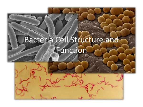
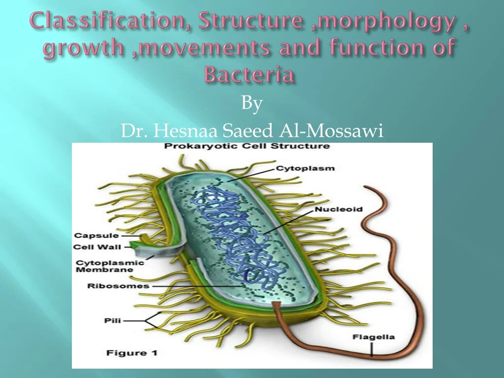
Classification, Structure ,morphology , growth ,movements and function of Bacteria By Dr. Hesnaa Saeed Al-Mossawi
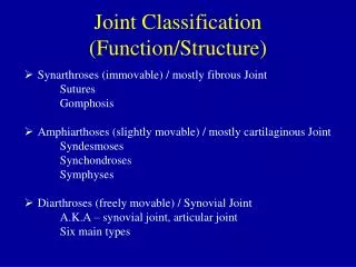
Classification • Kingdom of monera is made up of simple single celled organisms. • Prokaryotic cells • Monerans’ have four Phyla • Phylum: Schizophyta- The largest phylum of Monera. Contains organisms commonly known as bacteria • Phylum: Archaebateria- primitive organisms that live in harsh climates • Phylum: Cyanophyta: Organisms known as blue-green bacteria • Phylum:Prochlorophyta: Monera that live in marine environments
Growth • It is an increase in all the cell components, which ends in multiplication of cell leading to an increase in population. • It involves - an increase in the size of the cell & an increase in the number of individual cells. • Bacteria divide by binary fission.
Structure of Bacteria The cellular world is divided into two major groups, based on whether or not the cells have a nucleus (that is, an internal membrane-enclosed region that contains the genetic material). Cells that have a well-defined nucleus are called eukaryotic, whereas cells that lack a nucleus are called prokaryotic. All bacteria are prokaryotes. Size of Bacteria Average bacteria 0.5 - 2.0 um in diam. Surface Area ~12 um^2 Volume is ~4 um Surface Area to Volume is 3:1
Shapes of Bacteria • Coccus • Chain = Streptococcus • Cluster = Staphylococcus • Bacillus • Chain = Streptobacillus • Coccobacillus • Vibrio = curved • Spirillum • Spirochete • Square • Star Arrangements Clusters, tetrads, pairs, chains
Bacterial Structures • Flagella • Pili • Cell envelope • Cell membrane • peptidoglycan • Capsule • Gram + • Gram- • Cytoplasm • Cell Wall • Spores
Flagella • Flagella: Prokaryotic flagella are long, semirigid, helical, hollow tubular structures composed of several thousand molecules of the protein flagellin. They enable bacteria to move in a directed fashion • Motility - movement • Swarming occurs with some bacteria • Spread across Petri Dish • Proteus species most evident • Arrangement basis for classification • Monotrichous; 1 flagella • Lophotrichous; tuft at one end • Amphitrichous; both ends • Peritrichous; all around bacteria
Pili • Short protein appendages • smaller than flagella • Adhere bacteria to surfaces • F-pilus; used in conjugation • Exchange of genetic information • Flotation; increase buoyancy • Pellicle (scum on water)
THE CELL ENVELOPE • The bacterial “cell envelope” is a term applied to all material external to and enclosing the cytoplasm. It consists of several chemically and functionally distinct layers • The cell envelope also includes the capsule or glycocalyx, if present.
C/ D-Capsule , Glycocalyxor Slime Layer • Is a sticky, viscous material that forms an extracellular coating around the cell. The material is usually a polysaccharide. • If the material is tightly bound to the cell and has an organized structure, it is called a capsule • If the material is loosely bound and amorphous, it is called a slime layer, or glycocalyx. • It allow cells to adhere to surfaces, protect bacteria from antibodies and phagocytosis, and act as diffusion barriers against some antibiotics and protect bacteria against dessication, or drying
Cell envelope B-peptidoglycan • Peptido-glycan Polymer (amino acids + sugars) • Unique to bacteria • Sugars; NAG & NAM • N-acetylglucosamine • N-acetymuramic acid • D form of Amino acids used not L form • Amino acids cross link NAG & NAM
Gram-positive organisms: • Gram-positive bacteria have thick, multilayered, peptidoglycan cell walls that are exterior to the cytoplasmic membrane. • The peptidoglycan in most gram-positive species is covalently linked to teichoic acid, which is essentially a polymer of substituted glycerol units linked by phosphodiester bonds. • The teichoic acids are major cell surface antigens. Teichoic acids are integrated into the peptidoglycan layers but not tethered to the cytoplasmic membrane. • Lipoteichoicacids are lipid modified and integrated by this moiety into the outer leaflet of the cytoplasmic membrane.
Gram-negative organisms: • Gram-negative bacteria have a more complex cell wall structure composed of two membranes (an outer membrane and an inner, that is, cytoplasmic, membrane). • The two membranes are separated by the periplasmic space, which contains the peptidoglycan layer. The periplasmic space also contains degradative enzymes and transport proteins. • In contrast to gram-positive cells, the peptidoglycan layer of gram-negative cells is thin, and the cells are consequently more susceptible to physical damage. • The outer membrane is distinguished by the presence of embedded lipopolysaccharide.
Gram-negative organisms: • Lipid A consists of phosphorylated glucosamine disaccharide units to which are attached a number of long-chain fatty acids • The lipid portion (called lipid A) is imbedded in the membrane and is toxic to humans and animals. • Because lipid A is an integral part of the membrane, it is called endotoxin, as opposed to exotoxins, which are secreted substances. • Appearance of Colonies • Mucoid = Smooth (lots of LPS or capsule) • Dry = Rough (little LPS or capsule)
A. Cytoplasmic membrane The cell membrane is composed of phospholipid, the molecules of which form two parallel surfaces (called a lipid bilayer) such that the polar phosphate groups are on the outside of the bilayer and the nonpolar lipid chains are on the inside. The membrane acts as a permeability barrier, restricting the kind and amount of molecules that enter and leave the cell.
Cytoplasm • 80% Water {20% Salts-Proteins) • Osmotic Shock important • DNA is single circular molecule, called the bacterial chromosome. The chromosome, along with several proteins and RNA molecules, forms an irregularly shaped structure called the nucleoid. Plasmids; extra circular DNA • Antibiotic Resistance • Ribosomes: structure made of RNA, site of protein synthesis • Inclusions: granules of sugar, lipid storage, etc. (storage) • No organelles (Mitochondria, Golgi, etc.)
Endospores • Resistant structure • Heat, irradiation, cold • Boiling >1 hr still viable • Takes time and energy to make spores • Location important in classification • Central, Subterminal, Terminal • Bacillus stearothermophilus -spores • Used for quality control of heat sterilization equipment • Bacillus anthracis - spores • Used in biological warfare