Download
1 / 50
560 likes | 827 Views
A comprehensive guide to the morphology of bacteria, including shapes, sizes, and arrangements, alongside detailed microscopy techniques such as light, phase contrast, fluorescent, and electron microscopy. Learn about staining techniques, bacterial cell wall structure, and functions of various cellular components.

E N D
EUKARYOTIC Parasites, fungi Membrane enclosed organelles Cytoskeleton MICROORGANISMS • PROKARYOTIC • Bacterial cell • Do not contain organelles • Cell wall, peptidoglycan
Prokaryote and eukaryote cells PROKARYOTE AND EUKARYOTE
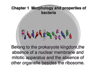
SIZE - 0.25-1 µm width 1-3 µm length SHAPE – cocci bacilli coccobacilli fusiform spiral ARRANGEMENT MORPHOLOGY OF BACTERIA
SHAPE OF BACTERIA Shapes of bacteria: 1. coccus; 2. bacillus; 3. vibrio; 4. spirillum; 5. spirochete
ARRANGEMENT OF BACTERIA Arrangement of curved bacteria: 1. vibrio; 2. spirilla; 3. spirochetes
OPTICAL/LIGHT MICROSCOPE MICROSCOPY Principle of bright-field (light) microscopy
PHASE CONTRAST MICROSCOPE Improves contrast Makes evident structures within cells ‘Phase differences’ converted to differences in intensity of light – producing light and dark contrast in image MICROSCOPY
FLUORESCENT MICROSCOPE Light of high intensity source excites a fluorescent agent, which in turn emits a low energy light of longer wavelength that produces the image MICROSCOPY
Fluorescent microscopy FLUORESCENT MICROSCOPE
FLUORESCENT MICROSCOPE APPLICATIONS Principles of fluorochroming and immunofluoresence
MICROSCOPY DARK GROUND MICROSCOPE Improves contrast Reflected light used instead of transmitted light Dark field condenser Light rays falling on the object are reflected or scattered on to objective lens Object self-luminous against dark background
DARK GROUND MICROSCOPE Dark-field microscopy
ELECTRON MICROSCOPE Beam of electrons instead of beam of light Electron beam focused by circular electromagnets - analogous to lens in light microscope Object held in the path of electron beam and produces an image which is focused on a screen MICROSCOPY
Shadow casting Negative staining – phosphotungstic acid Freeze etching ELECTRON MICROSCOPE APPLICATIONS
SIMPLE STAIN – Methylene blue or basic fuchsin NEGATIVE STAINING – India ink or nigrosin IMPREGNATION – Silver impregnation STAINING TECHNIQUES
Differential stains GRAM STAIN – Christian Gram (1884) Principle: Gram positive – acidic protoplasm - retain basic primary dye Peptidoglycan of Gram-positive bacteria thick – retain the dye iodine complex High lipid content of Gram-negative bacteria makes them permeable to counterstain (secondary dye) STAINING TECHNIQUES
ACID FAST STAIN - Ehrlich Modified – ZIEHL and NEELSEN Principle: High lipid content, variety of fatty acids and lipids Mycolic acid – peculiar to acid fast bacilli Integrity of the cell wall STAINING TECHNIQUES
ALBERT STAIN - demonstrate metachromatic granules – C.diphtheriae Neisser’s stain Ponder’s stain STAINING TECHNIQUES
BACTERIAL CELL Diagram of an idealised bacterial cell
Cell envelope and appendages Cell interior Cell structures function as a complex integrated unit BACTERIAL CELL
CELL ENVELOPE Outer membrane (GNB) Cell wall – peptidoglycan Periplasm (GNB) Cytoplasmic membrane BACTERIAL CELL
Bilayered Composed of lipopolysaccharide (LPS) Gives GNB - net negative charge Scattered throughout LPS - porins Porins - control passage of nutrients and antibiotics OUTER MEMBRANE
Composed of disaccharide-pentapeptide Disaccharide-N-acetyl glucosamine N-acetyl muramic acid Amino acid only linked to N-acetyl muramic acid Polymers-crosslink-via peptide bridges to form peptidoglycan sheets CELL WALL
CHEMICAL STUCTURE OF BACTERIAL CELL WALL Chemical structure of bacterial cell wall
Notable difference - Gram positive and Gram negative Gram positive-Peptidoglycan thick Teichoic acid Glycerol Ribitol phosphate Mycolic acid CELL WALL
CELL WALL Gram-positive call wall
CELL WALL Gram-negative cell wall
Cannot be seen by light microscope Cannot stain with simple stain Demonstration - Plasmolysis - hypertonic solution bacterial ghost - Micro dissection - Reaction with specific antibody - Differential staining - Electron microscope CELL WALL
Found in Gram-negative bacilli Inner surface of outer membrane Contains the peptidoglycan layer Helps secure nutrients Has enzymes that degrade macromolecules and detoxify antibiotics PERIPLASMIC SPACE
Present in Gram-positive and Gram-negative bacteria Deepest layer of cell envelope Heavily laced with proteins and enzymes vital to cell metabolism Cytoplasmic membrane functionally similar to eukaryotic cell organelles (Mitochondria, Golgi, Lysosomes) CYTOPLASMIC MEMBRANE
Transport of solutes Enzymes involved in cell synthesis Generation of chemical energy Cell motility Chromosomal segregation CYTOPLASMIC MEMBRANE FUNCTIONS
Capsule Fimbria-pili Flagella Role in causing infection Help identification in laboratory CELLULAR APPENDAGES
Immediately exterior to peptidoglycan Glycocalyx/Slime Protect bacteria from attack of cells of human defense mechanism Facilitates and maintains bacterial colonisation of biological surface Example: teeth, prosthetic heart valve CAPSULE
SIGNIFICANCE Virulence - Inhibit phagocytosis - Protect cell from lysozyme Permit adherence - Cell surface Example: implant, catheters Prevents cell from drying Toxicity to host cell Protects cell from bacteriophage CAPSULE
Covalently linked to cell wall No net charge - so do not bind to dyes Gram stain – clear halo around bacteria Demonstrated by negative staining - India ink - Nigrosin - Congo red Demonstrated immunologically - Quellung reaction CAPSULE
CAPSULE S.pneumoniae capsule seen by India ink staining
Serological typing - capsular antigen Detection of antigen - CSF, blood Example: S.pneumoniae - CSF Neisseria meningitidis Vaccine - capsular polysaccharide food antigen CAPSULE APPLICATIONS
Short, hair-like structures Proteinaceous-antigenic Protrude through cell wall Two types: common pili-adhesins, sex pili FIMBRIAE/PILI
SIGNIFICANCE Act as adhesins - bacteria colonise Receptor for bacteriophage Streptococcus pyogenes - M protein Virulence factor Some fimbriae - agglutinate RBCs FIMBRIAE/PILI
Complex structure Composed of protein flagellin Embedded in cell envelope Motility - survival FLAGELLA
Non-contractile Single protein subunit - flagellin Anchored to bacterial cytoplasmic membrane by disc-like structure FLAGELLA
Three parts: filament, hook, basal body Flagella attached to cell body by hook and basal body Hook and basal body embedded in envelope Basal body -1 set of rings-Gram positive 2 sets of rings-Gram negative FLAGELLA
FLAGELLA - STRUCTURE General structure of the flagellum of a Gram-negative bacterium
ARRANGEMENT OF FLAGELLA Types of flagellar arrangement: 1. monotrichous; 2. lophotrichous; 3. amphitrichous; 4. amphilophotrichous; 5. peritrichous fl agella
Detection of motility-Direct-hanging drop Phase contrast Dark ground Motility media Demonstration of flagella-Flagella stain Electron microscope Immunology FLAGELLA
Adverse physical and chemical conditions Nutrients scarce Metabolic and structural change Bacterial spores – endospores Favorable conditions endospore germinates Spores killed – autoclave, formaldehyde SPORE
BACTERIAL SPORE Diagrammatic representation of a bacterial spore: 1. germinal groove; 2. outer cortical layer; 3. cortex; 4. internal spore coat; 5. subcoat material; 6. outer spore coat; 7. cytoplasmic membrane; 8. cell wall primordium.
TYPES OF BACTERIAL SPORES Types of bacterial spores: 1. central, bulging; 2. subterminal, bulging; 3. terminal, spherical; 4. central, not bulging; 5. subterminal, not bulging; 6. terminal, oval
Pleomorphism – defective cell wall synthesis Involution forms – activity of autolytic enzymes L forms – Kleineberger and Nobel - aberrant morphological forms Lister Institute, London PLEOMORPHISM