Download
1 / 42

E N D
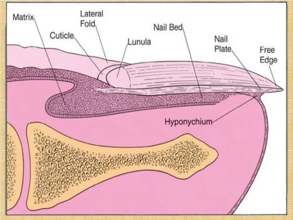
Effects of trauma Permanent ridges or splits in the nail plate can follow damage to the nail matrix. Splinter haemorrhages, the linear nature of which is determined by longitudinal ridges and grooves in the nail bed, are most commonly seen under the nails of manual workers and are caused by minor trauma. Larger subungualhaematomasare usually easy to identify and dark areas of altered blood can raise worries about the presence of a subungual melanoma. Chronic trauma from sport and from ill-fitting shoes contributes to haemorrhage under the nails of the big toes, to the gross thickening of toenails known as onychogryphosis and to ingrowing nails.
Onycholysis a separation of the nail plate from the nail bed, may be a result of minor trauma although it is also seen in nail psoriasis, phototoxic reactions, repeated immersion in water, after the use of nail hardeners and possibly in thyroid disease. Usually, no cause for it is found. The space created may be colonized by yeasts, or by bacteria such as Pseudomonas aeruginosa, which turns it an ugly green colour.
Some nervous habits damage the nails. Bitten nails are short and irregular; some people also bite their cuticles and the skin around the nails.Viral warts can be seeded rapidly in this way. In the common habit tic nail dystrophy, the cuticle of the thumbnail is the target for picking or rubbing.This repetitive trauma causes a ladder pattern of transverse ridges and grooves to run up the centre of the nail plate.
Lamellar splitting of the distal part of the fingernails, so commonly seen in housewives, has been attributed to repeated wetting and drying. Attempts to beautify nails can lead to contact allergy. Culprits include the acrylate adhesive used with artificial nails and formaldehyde in nail hardeners. In contrast, contact dermatitis caused by allergens in nail polish itself seldom affects the fingers but presents as small itchy eczematous areas where the nail plates rest against the skin during sleep. The eyelids, face and neck are favourite sites.
The nail in systemic disease Clubbing: A bulbous enlargement ofthe terminal phalanx with an increase in the angle between the nail plate and the proximal fold to over 180°. Its association with chronic lung disease and with cyanotic heart disease is well known. Rarely, clubbing may be familial with no underlying cause. The mechanisms involved in itsformation are still not known.
Koilonychia: A spooning and thinning of the nail plate, indicating iron deficiency Colour changes:The ‘half-and-half’ nail, with awhite proximal and red or brown distal half, is seen in a minority of patients with chronic renal failure. Whitening of the nail plates may be related to hypoalbuminaemia, as in cirrhosis of the liver. Some drugs, notably antimalarials, antibiotics and phenothiazines, can discolour the nails.
Beau’s lines:Transverse grooves that appear synchronously on all nails a few weeks after an acute illness, and which grow steadily out to the free margin . Connective tissue disorders:Nail fold telangiectasia or erythema is a useful physical sign in dermatomyositis,systemic sclerosis and systemic lupus erythematosus. In dermatomyositis, the cuticles become shaggy, and in systemic sclerosis loss of finger pulp leads to overcurvature of the nail plates. Thin nails, with longitudinal ridging and sometimes partial onycholysis, are seen when the peripheral circulation is impaired, as in Raynaud’s phenomenon.
Nail changes in the common dermatoses Psoriasis: severe nail involvement is more likely in the presence of arthritis. The best-known nail change is pitting of the surface of the nail plate .Almost as common is psoriasis under the nail plate, showing up as red or brown areas resembling oil spots, often with onycholysis bordered by obvious discoloration. Eczema: Some patients with itchy chronic eczema bring their nails to a high state of polish by scratching. In addition,eczema of the nail folds may lead to a coarse irregularity with transverse ridging of the adjacent nail plates.
Lichen planus: Most often this is a reversible thinning of the nail plate with irregular longitudinal grooves and ridges. More severe involvement may lead to pterygiumin which the cuticle grows forward over the base of the nail and attaches itself to the nailplate. The threat of severe and permanent nail changes can sometimes justify treatment with systemic steroids. Alopecia areata: The more severe the hair loss, the more likely there is to be nail involvement. A roughness or fine pitting is seen on the surface of the nail plates and the lunulae may appear mottled.
Infections Acute paronychia: The portal of entry for the organisms concerned,usually staphylococci, is a break in the skin or cuticle as a result of minor trauma. The subsequent acute inflammation, often with the formation of pus in the nail fold or under the nail, requires systemic treatment with flucloxacillin, cephalexinorerythromycin and appropriate surgical drainage.
Chronic paronychia: A combination of circumstances can allow a mixture of opportunistic pathogens (yeasts, Gram-positivecocci and Gram-negative rods) to colonize the space between the nail fold and nail plate, producing a chronic dermatitis. Predisposing factors include a poor peripheral circulation, wet work, working with flour, diabetes, vaginal candidosis and overvigorous cutting back of the cuticles.
The nail folds become tender and swollen and small amounts of pus are discharged at intervals. The cuticular seal is damaged and the adjacent nail plate becomes ridged and discoloured. Paronychia should not be confused with a dermatophyte infection in which the nail folds are not primarily affected. Manicuring of the cuticle should cease. The hands should be kept as warm and as dry as possible, and the damaged nailfolds packed several times a day with an imidazole cream. Highly potent topical corticosteroid creams applied for 3 weeks also help. If there is no response, and swabs confirm that Candida is present, a 2-week course of itraconazole should be considered.
Dermatophyte infections The common dermatophytes that cause tineapedis can also invade the nails. Toe nail infection is common and associated with tineapedis. The early changes often occur at the free edge of the nail and spread proximally. The nail plate becomes yellow, crumbly and thickened.Usually, only a few nails are infected but occasionally all are. The finger nails are involved less often and the changes, in contrast to those of psoriasis,are usually confined to one hand. Coexisting tineapedisfavoursdermatophyte infection of the nail. teatment include avoidance of the aggrevatings factors and intiation of systemic antifungals such as fluconazole 150mg once weekly or itaconazole 200mg twice daily for one week (pulse therapy)
Tumours Periungual warts are common and stubborn. Cryotherapy must be used carefully to avoid damage to the nail matrix, but is painful. Periungualfibromasarise from the nail folds, usually in late childhood, in patients with tuberous sclerosis. Glomustumourscan occur beneath the nail plate.The small red or bluish lesions are exquisitely painful if touched and when the temperature changes.Treatment is surgical. Subungualexostosesprotrude painfully under the nail plate. Usually secondary to trauma to the terminal phalanx, the bony abnormality can beseen on X-ray and treatment is surgical.
Myxoid cystsoccur on the proximal nail folds, usually of the fingers. The smooth domed swelling contains a clear jelly-like material that transilluminates well. A groove may form on the adjacent nail plate. Treatment by Cryotherapy, injections of triamcinolone and surgical excision . Malignant melanomashould be suspected in any subungual pigmented lesion, particularly if the pigment spreads to the surrounding skin (Hutchinson’ssign). Subungualhaematomas may cause confusion but ‘grow out’ with the nail .
Congenital syndromes nail–patella syndromethe thumb nails,and to a lesser extent those of the fingers, are smaller than normal. Rudimentary patellae, renal disease and iliac spines complete the syndrome, which is inherited as an autosomal dominant trait linked with the locus controlling ABO blood groups. Pachyonychiacongenitais also rare and inherited as an autosomal dominant trait. The nails are grossly thickened, especially peripherally, and have a curious triangular profile. Hyperkeratosis may occur on areas of friction on the legs and feet. EpidermolysisbullosaPermanent loss of the nails may be seen with the dystrophic types of Epidermolysisbullosa.
yellow nail syndromethe nail changes begin in adult life, against a background of hypoplasia of the lymphatic system. Peripheral oedema is usually present and pleural effusions may occur. The nails grow very slowly and become thickened and greenish-yellow; their surface is smooth but they are overcurved from side to side. The nail ‘en racquette’is a short broad nail, usually a thumbnail, which is seen insome 1–2% of the population and inherited as anautosomal dominant trait. The basic abnormality is shortness of the underlying terminal phalanx.