Download
1 / 25
250 likes | 291 Views
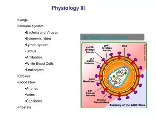
Understand the intricacies of cardiac and cerebral blood flow, coronary circulation, and blood vessel functions in cardiovascular physiology. Learn about the Fick Principle, endothelial factors, and the impact of NO and ET-1 on vascular dynamics. Gain insights into vessel classifications, from elastic arteries to capillaries.

E N D
Cardiac Physiology(III) A. Rüçhan Akar Ankara University School of Medicine December- 2003
Coronary Blood Flow • coronary blood flow: 250 ml/min • 5% of resting cardiac output • 60-80 ml blood/100g tissue/min • entirely during diastole ~ aortic diastolic pressure minus LVDP ~ duration of diastole • pressure < 150 mmHg • oxygenated by superb membrane oxygenator-”the lungs”
Cerebral Blood Flow • Cerebral blood flow: 750 ml/min • 15% of resting cardiac output • 50-55 ml blood/100g tissue/min
* The peak left coronary flow occurs at the end of isovolumetric relaxation * Left coronary blood flow Right coronary blood flow
mitochondria cellular pO2 < 5mmHg within seconds oxidative phosporilation stops cytosol anaerobic glycolysis glycogen glucose-6-phosphate pyruvate lactate cellular acidosis depletion of ATP Cessation of Myocardial Blood Flow
Depletion of ATP < 50% of Normal Level- irreversible lethal cell injury • glycolysis is blocked • increasing cellular acidity • protein denaturation • structural, enzymatic, nuclear changes
Measurement of Cardiac Output Fick Principle
The Fick Principle . VO2 CaO2 – CvO2 Q = – Q: cardiac output VO2: O2 consumption CaO2:arterial O2 content CvO2: mixed venous O2 content
Blood Vessel • Intima primarily the endothelial lining • Media vascular smooth muscle, collagen, elastin • Adventitia connective tissue
Vascular Endothelium Vasodilators Vasoconstrictors Nitric Oxide Prostacyclin Endothelium-derived hyperpolarizing factor Bradykinin Endothelin-1 Angiotensin II Wilson SH, Lerman A. Heart Physiology and Pathophysiology, Academic Press (edited by Sperelakis N.) 473-480
L-Arginine is converted to NO by the enzyme nitric oxide synthase (NOS)
Nitric Oxide (NO)Function • Vasodilator • Inhibitor of vascular smooth muscle cell proliferation • Inhibitor of platelet adherence/aggregation • Inhibitor of leukocyte/endothelial interactions
Endothelin-1(ET-1) • Peptide first sequenced in 1988 • Most potent vasoconstrictor in humans • Maintenance of basal arterial vasomotor tone • Strong chemoattractant for circulating monocytes and macrophage activation “proatherogenic”
Endothelial Dysfunction • Imbalance of endothelium-derived relaxing and contracting factors Atherosclerotic risk factors Decreased NO bioavailability Increased levels of ET-1
Functional Classification of Vessel Wall • elastic arteries • muscular arteries • resistance vessels • capillaries (exchange vessels) • venules (capacitance vessels) JR Levick, 1995 An Introduction to Cardiovascular Physiology Butterworth-Heinemann
Elastic Arteries • aorta, pulmonary artery and major branches • diameter = 1-2 cm • tunica media is rich in elastin (extensible) • collagen (prevents overdistension)
intima AORTA Elastic fibers Smooth muscle media adventitia Bergman RA, Afifi AK, Heidger PM Atlas of Microscopic Anatomy, 1989 W.B. Saunders Company
Conduit (Muscular) Arteries • diameter = 1mm-1cm • popliteal, radial, cerebral, coronary arteries • tunica media is thicker, contains more smooth muscle • rich autonomic nerve supply (contraction and relaxation)
Resistance Vessels • main resistance to blood flow resides in the; • smallest, terminal arteries (diameter = 100-500mm) • arterioles (< 100mm) “single layer of muscle in the media” • richly innervated by vasoconstrictor nerve fibres • actively regulate local blood flow to match local demand
Capillaries (Exchange vessels) • diameter: 4-7mm • wall: single layer of endothelial cells • wall thickness = 0.5mm • large cross-sectional area • slow blood velocity • red cell transit time = 1-2 sec
Arteriovenous Anastomosis • shunt vessels ( diameter = 20-135mm) • connect arterioles to venules, bypassing the capillaries • skin, nasal mucosa • temperature regulation
The Veins“Capacitance Vessels” • diameter 50-200mm • thin wall • in limb veins, intima possesses pairs of valves • low resistance to flow • storing large volumes of blood under low pressure • ~ 60-70% of the circulating blood volume
Distribution of blood volume in a resting man ( 5.5 litres) Folkow B, Neil E. 1971, Oxford University Press, London