Download
1 / 1
10 likes | 179 Views
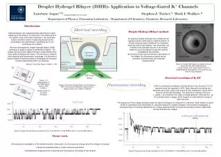
Droplet Hydrogel Bilayer (DHB)- Application to Voltage-Gated K + Channels Lauriane Angué 1,2 l.angue1@physics.ox.ac.uk Stephen J. Tucker 1 / Mark I. Wallace 2 Department of Physics, Clarendon Laboratory / Department of Chemistry, Chemistry Research Laboratory. Introduction:

E N D
Droplet Hydrogel Bilayer (DHB)- Application to Voltage-Gated K+ Channels Lauriane Angué 1,2l.angue1@physics.ox.ac.ukStephen J. Tucker 1/ Mark I. Wallace 2 Department of Physics, Clarendon Laboratory / Department of Chemistry, Chemistry Research Laboratory Introduction: Artificial bilayers are a supplementary alternative to patch-clamping for the study of ion channels. The components of the system, such as the lipid compostion, the electrolyte solution and the number of channels studied can be controlled by the experimenter; something the in vivo techniques do not allow. We have developed the droplet-hydrogel bilayer (DHB) technique to study a variety of membrane proteins*. The bilayer formed is quite stable and has a longer lifetime than other artificial bilayers techniques. This technique combines both electrical and fluorescence recordings. It is therefore a usefull method if someone wants to investigate the structure-function relationships within ion channels. * Bayley H. et al, Mol. Biosyst. (2008) 4, 1191. Electrical recording Droplet Hydrogel Bilayer method: An aqueous droplet is brought into contact with an agarose surface immersed in a lipid/oil solution. A droplet hydrogel bilayer (DHB) will form between these two with a high stability. Two electrodes, one inserted in the hydrogel and one in the droplet detect the ion flux passing through the channel embedded at the interface. By directing a total internal reflection laser beam on the bilayer, the fluorescence signal can be recorded, simultaneously with electrical measurements. Figure 3: A bright-field image of a droplet forming a bilayer with the lipid-coated agarose. Image courtesy of Dr. Lydia Harriss Thesis “A Single Molecule Investigation of Ion Channels and Pore-Forming Peptides” Electrical recordings of KvAP: KvAP is a prokaryotic potassium channel whose X-ray structure of 3.2 Å resolution was first reported in 2003. Each channel is formed by four identical subununits, which form a pore at their intersection, where the K+ ions can go through the bilayer. The opening or closing of the pore (« gating ») is controled in the case of voltage-gated K+ channels (Kv), by the transmembrane voltage, which triggers a conformational change to open the pore. Figure 1: A representation of the PMMA device used for DHB formation. Image courtesy of Dr James Thompson Fluorescence recording Figure 2: A droplet hydrogel bilayer for simultaneous TIRF microscopy and electrical measurements. Image courtesy of Dr James Thompson KvAP-C247S The sequence of this voltage-sensing domain is highly homologous to eukaryotic Kv channels, which makes it a good model to understand how mammalian Kv channels respond to voltage changes. In the present investigation, a fluorescent dye is covalently attached to the voltage sensing domain to probe the structure-function relationship through activation by voltage. KvAP-L121C-C247S labelled with TMRM Figure 4: Single channel traces recorded at 0 mV in 150 mM KCl, 10 mM HEPES, pH=7.0, by the DHB method. • Future work: • Fluorescence investigation of the labelled mutants: observation of a fluorescence change when the voltage is changed. • Scale the quantities down to single molecule experiment. • Simultaneous measurements of electrical and fluorescence recordings of the mutants. Figure 5: Single channel traces recorded at 0 mV in 150 mM KCl, 10 mM HEPES, pH=7.0, by the DHB method.