Understanding DNA Structure and the Role of Restriction Enzymes
120 likes | 246 Views
This overview explores the basic structure of DNA, highlighting its composition from nucleotides, including pentose sugar, phosphate, and nitrogen bases (adenine, guanine, thymine, and cytosine). DNA's double helix formation, according to Rosalind Franklin's findings, operates on Chargaff’s Rule for base pairing. We delve into the mechanisms of restriction enzymes, their bacterial origins, and how they cut DNA at specific sequences, producing blunt or sticky ends. Additionally, the process of gel electrophoresis for separating DNA fragments by size is discussed, along with its importance in genetic analysis.

Understanding DNA Structure and the Role of Restriction Enzymes
E N D
Presentation Transcript
Basic Structure of DNA • Built of nucleotides: • Pentose sugar • Phosphate • Nitrogen base • Purines – adenine, guanine • Pyrmidines – thymine, cytosine • N base attached to 1’ C of sugar
Struct., cont. • 3’ C of 1 sugar bonds to 5’ phosphate to form phosphodiester bond
Complementary Strands • DNA arranged in double helix (Rosalind Franklin’s work) • Chargaff’s Rule: A—T and G—C (purine to pyrimidine) • Antiparallel – run 5’3’ on 1 strand and 3’5’ on other • 2 strands are complementary; i.e. • 3’—AGTAC—5’ • 5’—TCATG—3’
Properties of DNA • Negative charge • PO4- • Together, negative charge and polar sugar = soluble • Genes & non-coding sections • DNA is universal
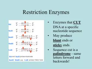
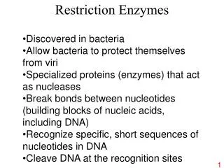
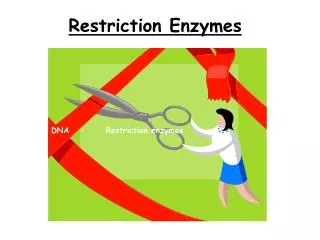
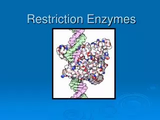
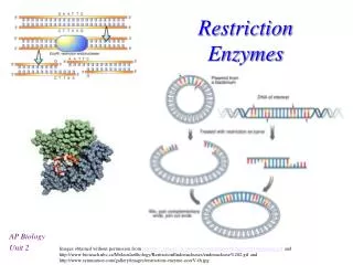
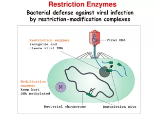
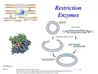
Restriction Enzymes • Isolated from bacteria • Used as immune mechanism to protect from phages • Named after bacteria they are isolated from • Recognize specific sequences of DNA • Palindromes • Ex. EcoRI recognizes GAATTC
“Cut” DNA in a specific pattern at the recognition site • May produce: • Blunt ends • Sticky ends
Number & placement of recognition sites determine # and size of fragments • RFLPs • http://highered.mcgraw-hill.com/sites/0072437316/student_view0/chapter16/animations.html#
Gel Electrophoresis • Separates DNA molecules based on size • Cut DNA using restriction enzyme(s) • Load DNA into gel • Run electrical current through buffer/gel • Stain DNA & compare unknowns to fragments of known sizes